6HRN

C-Phycocyanin from heterocyst forming filamentous cyanobacterium Nostoc sp. WR13


Experimental Data Snapshot

  • Method: X-RAY DIFFRACTION
  • Resolution: 1.51 Å
  • R-Value Free: 0.154 
  • R-Value Work: 0.115 
  • R-Value Observed: 0.117 

wwPDB Validation   3D Report Full Report


Ligand Structure Quality Assessment 


This is version 1.1 of the entry. See complete history


Literature

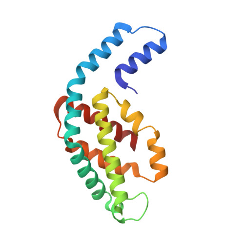
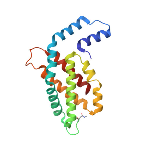
Crystal structure of phycocyanin from heterocyst-forming filamentous cyanobacterium Nostoc sp. WR13.

Patel, H.M.Roszak, A.W.Madamwar, D.Cogdell, R.J.

(2019) Int J Biol Macromol 135: 62-68

  • DOI: https://doi.org/10.1016/j.ijbiomac.2019.05.099
  • Primary Citation of Related Structures:  
    6HRN

  • PubMed Abstract: 

    Phycocyanin (PC) is the principal pigment protein in the light-harvesting antenna of cyanobacteria. Here the biochemical characterization and the 1.51 Å crystal structure of PC from cyanobacterium Nostoc sp. WR13 (Nst-PC) is reported. The P6 3 crystal lattice is composed of the minimal biological entities of Nst-PC, the (αβ) 3 trimeric rings. The structure has been refined to R factor 11.5% (R free 15.4%) using anisotropic atomic B factors. A phylogenetic study shows that the α and β chains of Nst-PC are significantly clustered in a distinct clade with Acaryochloris marina. The structure was examined to look for any significant differences between Nst-PC and PC from non-desert species. Only minor differences were found in the chromophore microenvironments. The tentative energy transfer pathways in Nst-PC were modeled based on simple structural considerations.


  • Organizational Affiliation

    P. G. Department of Biosciences, Satellite Campus, Vadtal Road, Sardar Patel University, Bakrol, 388315 Anand, Gujarat, India; Institute of Molecular Cell and Systems Biology, University of Glasgow, 120 University Place, Glasgow G12 8TA, UK. Electronic address: hiralpatel2387@yahoo.co.in.


Macromolecules
Find similar proteins by:  (by identity cutoff)  |  3D Structure
Entity ID: 1
MoleculeChains Sequence LengthOrganismDetailsImage
Alpha Subunit of Cyanobacterial Phycocyanin protein162Nostoc sp.Mutation(s): 0 
UniProt
Find proteins for A0A509GUZ8 (Nostoc sp)
Explore A0A509GUZ8 
Go to UniProtKB:  A0A509GUZ8
Entity Groups  
Sequence Clusters30% Identity50% Identity70% Identity90% Identity95% Identity100% Identity
UniProt GroupA0A509GUZ8
Sequence Annotations
Expand
  • Reference Sequence
Find similar proteins by:  (by identity cutoff)  |  3D Structure
Entity ID: 2
MoleculeChains Sequence LengthOrganismDetailsImage
Beta Subunit of Cyanobacterial Phycocyanin protein172Nostoc sp.Mutation(s): 0 
UniProt
Find proteins for A0A509GUZ9 (Nostoc sp)
Explore A0A509GUZ9 
Go to UniProtKB:  A0A509GUZ9
Entity Groups  
Sequence Clusters30% Identity50% Identity70% Identity90% Identity95% Identity100% Identity
UniProt GroupA0A509GUZ9
Sequence Annotations
Expand
  • Reference Sequence
Small Molecules
Ligands 7 Unique
IDChains Name / Formula / InChI Key2D Diagram3D Interactions
CYC
Query on CYC

Download Ideal Coordinates CCD File 
C [auth A],
F [auth B],
G [auth B]
PHYCOCYANOBILIN
C33 H40 N4 O6
VXTXPYZGDQPMHK-GMXXPEQVSA-N
PG4
Query on PG4

Download Ideal Coordinates CCD File 
D [auth A],
I [auth B],
J [auth B],
K [auth B],
L [auth B]
TETRAETHYLENE GLYCOL
C8 H18 O5
UWHCKJMYHZGTIT-UHFFFAOYSA-N
PGE
Query on PGE

Download Ideal Coordinates CCD File 
N [auth B],
O [auth B]
TRIETHYLENE GLYCOL
C6 H14 O4
ZIBGPFATKBEMQZ-UHFFFAOYSA-N
MPD
Query on MPD

Download Ideal Coordinates CCD File 
Q [auth B](4S)-2-METHYL-2,4-PENTANEDIOL
C6 H14 O2
SVTBMSDMJJWYQN-YFKPBYRVSA-N
PEG
Query on PEG

Download Ideal Coordinates CCD File 
E [auth A],
M [auth B]
DI(HYDROXYETHYL)ETHER
C4 H10 O3
MTHSVFCYNBDYFN-UHFFFAOYSA-N
PO4
Query on PO4

Download Ideal Coordinates CCD File 
H [auth B]PHOSPHATE ION
O4 P
NBIIXXVUZAFLBC-UHFFFAOYSA-K
NO3
Query on NO3

Download Ideal Coordinates CCD File 
P [auth B]NITRATE ION
N O3
NHNBFGGVMKEFGY-UHFFFAOYSA-N
Modified Residues  1 Unique
IDChains TypeFormula2D DiagramParent
MEN
Query on MEN
B
L-PEPTIDE LINKINGC5 H10 N2 O3ASN
Experimental Data & Validation

Experimental Data

  • Method: X-RAY DIFFRACTION
  • Resolution: 1.51 Å
  • R-Value Free: 0.154 
  • R-Value Work: 0.115 
  • R-Value Observed: 0.117 
  • Space Group: P 63
Unit Cell:
Length ( Å )Angle ( ˚ )
a = 151.473α = 90
b = 151.473β = 90
c = 39.764γ = 120
Software Package:
Software NamePurpose
REFMACrefinement
XDSdata reduction
pointlessdata scaling
Aimlessdata scaling
AutoPROCdata scaling
STARANISOdata scaling
PHASERphasing

Structure Validation

View Full Validation Report


Ligand Structure Quality Assessment 


Entry History 

Deposition Data

Revision History  (Full details and data files)

  • Version 1.0: 2019-06-05
    Type: Initial release
  • Version 1.1: 2019-06-12
    Changes: Data collection, Database references