6HPC

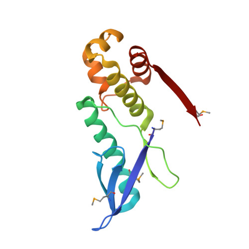
Crystal structure of the HicB antitoxin from E. coli


Experimental Data Snapshot

  • Method: X-RAY DIFFRACTION
  • Resolution: 2.26 Å
  • R-Value Free: 0.268 
  • R-Value Work: 0.227 

wwPDB Validation   3D Report Full Report


This is version 1.2 of the entry. See complete history


Literature

The E. coli HicB Antitoxin Contains a Structurally Stable Helix-Turn-Helix DNA Binding Domain.

Manav, M.C.Turnbull, K.J.Jurenas, D.Garcia-Pino, A.Gerdes, K.Brodersen, D.E.

(2019) Structure 27: 1675-1685.e3

  • DOI: https://doi.org/10.1016/j.str.2019.08.008
  • Primary Citation of Related Structures:  
    6HPB, 6HPC

  • PubMed Abstract: 

    The E. coli hicAB type II toxin-antitoxin locus is unusual by being controlled by two promoters and by having the toxin encoded upstream of the antitoxin. HicA toxins contain a double-stranded RNA-binding fold and cleaves both mRNA and tmRNA in vivo, while HicB antitoxins contain a partial RNase H fold and either a helix-turn-helix (HTH) or ribbon-helix-helix domain. It is not known how an HTH DNA-binding domain affects higher-order structure for the HicAB modules. Here, we present crystal structures of the isolated E. coli HicB antitoxin and full-length HicAB complex showing that HicB forms a stable DNA-binding module and interacts in a canonical way with HicA despite the presence of an HTH-type DNA-binding domain. No major structural rearrangements take place upon binding of the toxin. Both structures expose well-ordered DNA-binding motifs allowing a model for DNA binding by the antitoxin to be generated.


  • Organizational Affiliation

    Department of Molecular Biology and Genetics, Aarhus University, Centre for Bacterial Stress Response and Persistence, Aarhus 8000, Denmark.


Macromolecules
Find similar proteins by:  (by identity cutoff)  |  3D Structure
Entity ID: 1
MoleculeChains Sequence LengthOrganismDetailsImage
Antitoxin HicBA [auth B],
B [auth A]
138Escherichia coli 2-210-07_S3_C3Mutation(s): 0 
Gene Names: hicBAC45_3823
UniProt
Find proteins for P67697 (Escherichia coli (strain K12))
Explore P67697 
Go to UniProtKB:  P67697
Entity Groups  
Sequence Clusters30% Identity50% Identity70% Identity90% Identity95% Identity100% Identity
UniProt GroupP67697
Sequence Annotations
Expand
  • Reference Sequence
Small Molecules
Modified Residues  1 Unique
IDChains TypeFormula2D DiagramParent
MSE
Query on MSE
A [auth B],
B [auth A]
L-PEPTIDE LINKINGC5 H11 N O2 SeMET
Experimental Data & Validation

Experimental Data

  • Method: X-RAY DIFFRACTION
  • Resolution: 2.26 Å
  • R-Value Free: 0.268 
  • R-Value Work: 0.227 
  • Space Group: P 1 21 1
Unit Cell:
Length ( Å )Angle ( ˚ )
a = 43.09α = 90
b = 60.38β = 103.76
c = 59.34γ = 90
Software Package:
Software NamePurpose
XDSdata reduction
XSCALEdata scaling
CRANK2phasing
PHENIXrefinement

Structure Validation

View Full Validation Report


Entry History & Funding Information

Deposition Data


Funding OrganizationLocationGrant Number
Novo Nordisk FoundationDenmarkNNF17OC0030646
Danish National Research FoundationDenmarkDNRF120

Revision History  (Full details and data files)

  • Version 1.0: 2019-09-18
    Type: Initial release
  • Version 1.1: 2019-10-16
    Changes: Data collection
  • Version 1.2: 2019-11-13
    Changes: Database references