6HMY

Cholera toxin classical B-pentamer in complex with fucosyl-GM1


Experimental Data Snapshot

  • Method: X-RAY DIFFRACTION
  • Resolution: 1.60 Å
  • R-Value Free: 0.214 
  • R-Value Work: 0.184 
  • R-Value Observed: 0.185 

wwPDB Validation   3D Report Full Report


Ligand Structure Quality Assessment 


This is version 2.1 of the entry. See complete history


Literature

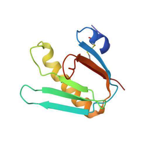
Crystal structures of cholera toxin in complex with fucosylated receptors point to importance of secondary binding site.

Heim, J.B.Hodnik, V.Heggelund, J.E.Anderluh, G.Krengel, U.

(2019) Sci Rep 9: 12243-12243

  • DOI: https://doi.org/10.1038/s41598-019-48579-2
  • Primary Citation of Related Structures:  
    6HJD, 6HMW, 6HMY

  • PubMed Abstract: 

    Cholera is a life-threatening diarrhoeal disease caused by the human pathogen Vibrio cholerae. Infection occurs after ingestion of the bacteria, which colonize the human small intestine and secrete their major virulence factor - the cholera toxin (CT). The GM1 ganglioside is considered the primary receptor of the CT, but recent studies suggest that also fucosylated receptors such as histo-blood group antigens are important for cellular uptake and toxicity. Recently, a special focus has been on the histo-blood group antigen Lewis x (Le x ), however, where and how the CT binds to Le x remains unclear. Here we report the high-resolution crystal structure (1.5 Å) of the receptor-binding B-subunits of the CT bound to the Le x trisaccharide, and complementary quantitative binding data for CT holotoxins. Le x , and also L-fucose alone, bind to the secondary binding site of the toxin, distinct from the GM1 binding site. In contrast, fucosyl-GM1 mainly binds to the primary binding site due to high-affinity interactions of its GM1 core. Le x is the first histo-blood group antigen of non-secretor phenotype structurally investigated in complex with CT. Together with the quantitative binding data, this allows unique insight into why individuals with non-secretor phenotype are more prone to severe cholera than so-called 'secretors'.


  • Organizational Affiliation

    Department of Chemistry, University of Oslo, P.O. Box 1033, NO-0315, Blindern, Norway.


Macromolecules
Find similar proteins by:  (by identity cutoff)  |  3D Structure
Entity ID: 1
MoleculeChains Sequence LengthOrganismDetailsImage
Cholera enterotoxin B-subunit
A, B, C, D, E
A, B, C, D, E, F, G, H, I, J
103Vibrio choleraeMutation(s): 0 
Gene Names: ctxBC9J66_18955EN12_07055ERS013165_03981ERS013197_06217ERS013202_03762ERS013206_03003
UniProt
Find proteins for P01556 (Vibrio cholerae serotype O1 (strain ATCC 39315 / El Tor Inaba N16961))
Explore P01556 
Go to UniProtKB:  P01556
Entity Groups  
Sequence Clusters30% Identity50% Identity70% Identity90% Identity95% Identity100% Identity
UniProt GroupP01556
Sequence Annotations
Expand
  • Reference Sequence
Oligosaccharides

Help

Entity ID: 2
MoleculeChains Length2D Diagram Glycosylation3D Interactions
alpha-L-fucopyranose-(1-2)-beta-D-galactopyranose-(1-3)-2-acetamido-2-deoxy-beta-D-galactopyranose-(1-4)-[N-acetyl-alpha-neuraminic acid-(2-3)]beta-D-galactopyranose-(1-4)-beta-D-glucopyranose
K, N, O, P, S
K, N, O, P, S, T
6N/A
Glycosylation Resources
GlyTouCan:  G91532KA
GlyCosmos:  G91532KA
GlyGen:  G91532KA
Entity ID: 3
MoleculeChains Length2D Diagram Glycosylation3D Interactions
alpha-L-fucopyranose-(1-2)-beta-D-galactopyranose-(1-3)-2-acetamido-2-deoxy-beta-D-galactopyranose-(1-4)-[N-acetyl-alpha-neuraminic acid-(2-3)]beta-D-galactopyranose-(1-4)-beta-D-glucopyranose
L, R
7N/A
Glycosylation Resources
GlyTouCan:  G91532KA
GlyCosmos:  G91532KA
GlyGen:  G91532KA
Entity ID: 4
MoleculeChains Length2D Diagram Glycosylation3D Interactions
alpha-L-fucopyranose-(1-2)-beta-D-galactopyranose-(1-3)-2-acetamido-2-deoxy-beta-D-galactopyranose-(1-4)-[N-acetyl-alpha-neuraminic acid-(2-3)]beta-D-galactopyranose-(1-4)-alpha-D-glucopyranose
M, Q
7N/A
Glycosylation Resources
GlyTouCan:  G18193SZ
GlyCosmos:  G18193SZ
GlyGen:  G18193SZ
Small Molecules
Ligands 3 Unique
IDChains Name / Formula / InChI Key2D Diagram3D Interactions
FUC
Query on FUC

Download Ideal Coordinates CCD File 
OA [auth F],
PA [auth J]
alpha-L-fucopyranose
C6 H12 O5
SHZGCJCMOBCMKK-SXUWKVJYSA-N
BCN
Query on BCN

Download Ideal Coordinates CCD File 
BA [auth B]
CA [auth C]
EA [auth C]
GA [auth D]
JA [auth D]
BA [auth B],
CA [auth C],
EA [auth C],
GA [auth D],
JA [auth D],
KA [auth E],
MA [auth E],
V [auth A],
W [auth A],
Y [auth B]
BICINE
C6 H13 N O4
FSVCELGFZIQNCK-UHFFFAOYSA-N
CA
Query on CA

Download Ideal Coordinates CCD File 
AA [auth B]
DA [auth C]
FA [auth C]
HA [auth D]
IA [auth D]
AA [auth B],
DA [auth C],
FA [auth C],
HA [auth D],
IA [auth D],
LA [auth E],
NA [auth E],
U [auth A],
X [auth A],
Z [auth B]
CALCIUM ION
Ca
BHPQYMZQTOCNFJ-UHFFFAOYSA-N
Experimental Data & Validation

Experimental Data

  • Method: X-RAY DIFFRACTION
  • Resolution: 1.60 Å
  • R-Value Free: 0.214 
  • R-Value Work: 0.184 
  • R-Value Observed: 0.185 
  • Space Group: C 1 2 1
Unit Cell:
Length ( Å )Angle ( ˚ )
a = 148.626α = 90
b = 74.137β = 105.59
c = 111.245γ = 90
Software Package:
Software NamePurpose
REFMACrefinement
Cootmodel building
XDSdata reduction
Aimlessdata scaling
PHASERphasing

Structure Validation

View Full Validation Report


Ligand Structure Quality Assessment 


Entry History & Funding Information

Deposition Data


Funding OrganizationLocationGrant Number
Research Council of NorwayNorway247730

Revision History  (Full details and data files)

  • Version 1.0: 2019-08-14
    Type: Initial release
  • Version 1.1: 2019-10-02
    Changes: Data collection, Database references
  • Version 2.0: 2020-07-29
    Type: Remediation
    Reason: Carbohydrate remediation
    Changes: Advisory, Atomic model, Data collection, Derived calculations, Refinement description, Structure summary
  • Version 2.1: 2024-01-24
    Changes: Data collection, Database references, Refinement description, Structure summary