Designed mono- and di-covalent inhibitors trap modeled functional motions for Trypanosoma cruzi proline racemase in crystallography.
Amaral, P.A., Autheman, D., de Melo, G.D., Gouault, N., Cupif, J.F., Goyard, S., Dutra, P., Coatnoan, N., Cosson, A., Monet, D., Saul, F., Haouz, A., Uriac, P., Blondel, A., Minoprio, P.(2018) PLoS Negl Trop Dis 12: e0006853-e0006853
- PubMed: 30372428 Search on PubMedSearch on PubMed Central
- DOI: https://doi.org/10.1371/journal.pntd.0006853
- Primary Citation Related Structures:
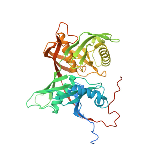
6HJE, 6HJF, 6HJG - PubMed Abstract:
Chagas disease, caused by Trypanosoma cruzi, affects millions of people in South America and no satisfactory therapy exists, especially for its life threatening chronic phase. We targeted the Proline Racemase of T. cruzi, which is present in all stages of the parasite life cycle, to discover new inhibitors against this disease. The first published crystal structures of the enzyme revealed that the catalytic site is too small to allow any relevant drug design. In previous work, to break through the chemical space afforded to virtual screening and drug design, we generated intermediate models between the open (ligand free) and closed (ligand bound) forms of the enzyme. In the present work, we co-crystallized the enzyme with the selected inhibitors and found that they were covalently bound to the catalytic cysteine residues in the active site, thus explaining why these compounds act as irreversible inhibitors. These results led us to the design of a novel, more potent specific inhibitor, NG-P27. Co-crystallization of this new inhibitor with the enzyme allowed us to confirm the predicted protein functional motions and further characterize the chemical mechanism. Hence, the catalytic Cys300 sulfur atom of the enzyme attacks the C2 carbon of the inhibitor in a coupled, regiospecific-stereospecific Michael reaction with trans-addition of a proton on the C3 carbon. Strikingly, the six different conformations of the catalytic site in the crystal structures reported in this work had key similarities to our intermediate models previously generated by inference of the protein functional motions. These crystal structures span a conformational interval covering roughly the first quarter of the opening mechanism, demonstrating the relevance of modeling approaches to break through chemical space in drug design.
- Université de Rennes 1, Equipe Chimie organique et interfaces (CORINT), UMR 6226 Sciences Chimiques de Rennes, Rennes, France.
Organizational Affiliation: