6HHK | pdb_00006hhk

Structure of gp105 of Listeria bacteriophage A511


Experimental Data Snapshot

  • Method: X-RAY DIFFRACTION
  • Resolution: 2.38 Å
  • R-Value Free: 
    0.243 (Depositor), 0.260 (DCC) 
  • R-Value Work: 
    0.204 (Depositor), 0.227 (DCC) 
  • R-Value Observed: 
    0.206 (Depositor) 

wwPDB Validation 3D Report Full Report

Validation slider image for 6HHK

This is version 1.2 of the entry. See complete history

Literature

Structure and transformation of bacteriophage A511 baseplate and tail upon infection ofListeriacells.

Guerrero-Ferreira, R.C.Hupfeld, M.Nazarov, S.Taylor, N.M.Shneider, M.M.Obbineni, J.M.Loessner, M.J.Ishikawa, T.Klumpp, J.Leiman, P.G.

(2019) EMBO J 38

  • DOI: https://doi.org/10.15252/embj.201899455
  • Primary Citation Related Structures: 
    6HHK

  • PubMed Abstract: 

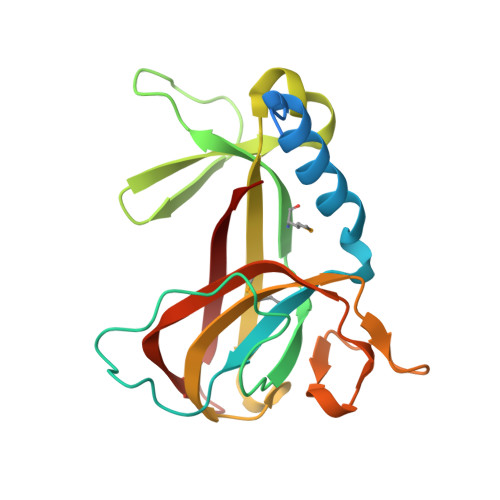
    Contractile injection systems (bacteriophage tails, type VI secretions system, R-type pyocins, etc.) utilize a rigid tube/contractile sheath assembly for breaching the envelope of bacterial and eukaryotic cells. Among contractile injection systems, bacteriophages that infect Gram-positive bacteria represent the least understood members. Here, we describe the structure of Listeria bacteriophage A511 tail in its pre- and post-host attachment states (extended and contracted, respectively) using cryo-electron microscopy, cryo-electron tomography, and X-ray crystallography. We show that the structure of the tube-baseplate complex of A511 is similar to that of phage T4, but the A511 baseplate is decorated with different receptor-binding proteins, which undergo a large structural transformation upon host attachment and switch the symmetry of the baseplate-tail fiber assembly from threefold to sixfold. For the first time under native conditions, we show that contraction of the phage tail sheath assembly starts at the baseplate and propagates through the sheath in a domino-like motion.


  • Organizational Affiliation
    • Laboratory of Structural Biology and Biophysics, School of Basic Sciences, École Polytechnique Fédérale de Lausanne, Lausanne, Switzerland.

Macromolecule Content 

  • Total Structure Weight: 64.48 kDa 
  • Atom Count: 4,187 
  • Modeled Residue Count: 526 
  • Deposited Residue Count: 579 
  • Unique protein chains: 1

Macromolecules

Find similar proteins by:|  3D Structure
Entity ID: 1
MoleculeChains  Sequence LengthOrganismDetailsImage
Gp105
A, B, C
193Pecentumvirus A511Mutation(s): 0 
UniProt
Find proteins for A8AST9 (Listeria phage A511)
Explore A8AST9 
Go to UniProtKB:  A8AST9
Entity Groups
Sequence Clusters30% Identity50% Identity70% Identity90% Identity95% Identity100% Identity
UniProt GroupA8AST9
Sequence Annotations
Expand
Reference Sequence

Small Molecules

Ligands 1 Unique
IDChains Name / Formula / InChI Key2D Diagram3D Interactions
CL

Query on CL


Download:Ideal Coordinates CCD File
D [auth A]CHLORIDE ION
Cl
VEXZGXHMUGYJMC-UHFFFAOYSA-M
Modified Residues  1 Unique
IDChains TypeFormula2D DiagramParent
MSE
Query on MSE
A, B, C
L-PEPTIDE LINKINGC5 H11 N O2 SeMET

Experimental Data & Validation

Experimental Data

  • Method: X-RAY DIFFRACTION
  • Resolution: 2.38 Å
  • R-Value Free:  0.243 (Depositor), 0.260 (DCC) 
  • R-Value Work:  0.204 (Depositor), 0.227 (DCC) 
  • R-Value Observed: 0.206 (Depositor) 
Space Group: P 32 2 1
Unit Cell:
Length ( Å )Angle ( ˚ )
a = 75.143α = 90
b = 75.143β = 90
c = 187.303γ = 120
Software Package:
Software NamePurpose
BUSTERrefinement
XDSdata reduction
XDSdata scaling
SHELXDEphasing

Structure Validation

View Full Validation Report


Entry History 

& Funding Information

Deposition Data


Funding OrganizationLocationGrant Number
Swiss National Science FoundationSwitzerland31003A_146284/1

Revision History  (Full details and data files)

  • Version 1.0: 2019-01-23
    Type: Initial release
  • Version 1.1: 2019-02-13
    Changes: Data collection, Database references
  • Version 1.2: 2024-11-20
    Changes: Data collection, Database references, Structure summary