6HGA | pdb_00006hga

Crystal Structure of the human IL-17RC D2-D3-D4 domains in complex with an anti-APP tag Fab


Experimental Data Snapshot

  • Method: X-RAY DIFFRACTION
  • Resolution: 2.60 Å
  • R-Value Free: 
    0.232 (Depositor) 
  • R-Value Work: 
    0.205 (Depositor) 
  • R-Value Observed: 
    0.206 (Depositor) 

wwPDB Validation 3D Report Full Report

Validation slider image for 6HGA

Ligand Structure Quality Assessment 


This is version 1.3 of the entry. See complete history

Literature

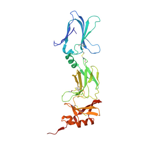
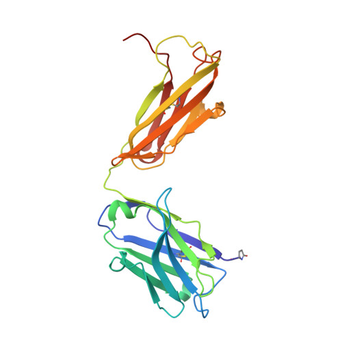
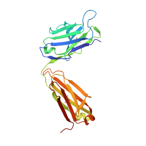
Structural Analysis Reveals that the Cytokine IL-17F Forms a Homodimeric Complex with Receptor IL-17RC to Drive IL-17RA-Independent Signaling.

Goepfert, A.Lehmann, S.Blank, J.Kolbinger, F.Rondeau, J.M.

(2020) Immunity 52: 499-512.e5

  • DOI: https://doi.org/10.1016/j.immuni.2020.02.004
  • Primary Citation Related Structures: 
    6HG4, 6HG9, 6HGA, 6HGO, 6HGU

  • PubMed Abstract: 

    Interleukin-17A (IL-17A), IL-17F, and IL-17A/F heterodimers are key cytokines of the innate and adaptive immune response. Dysregulation of the IL-17 pathway contributes to immune pathology, and it is therefore important to elucidate the molecular mechanisms that govern IL-17 recognition and signaling. The receptor IL-17RC is thought to act in concert with IL-17RA to transduce IL-17A-, IL-17F-, and IL-17A/F-mediated signals. We report the crystal structure of the extracellular domain of human IL-17RC in complex with IL-17F. In contrast to the expected model, we found that IL-17RC formed a symmetrical 2:1 complex with IL-17F, thus competing with IL-17RA for cytokine binding. Using biophysical techniques, we showed that IL-17A and IL-17A/F also form 2:1 complexes with IL-17RC, suggesting the possibility of IL-17RA-independent IL-17 signaling pathways. The crystal structure of the IL-17RC:IL-17F complex provides a structural basis for IL-17F signaling through IL-17RC, with potential therapeutic applications for respiratory allergy and inflammatory bowel diseases.


  • Organizational Affiliation
    • Novartis Institutes for BioMedical Research, Novartis Pharma AG, 4002 Basel, Switzerland.

Macromolecule Content 

  • Total Structure Weight: 78 kDa 
  • Atom Count: 5,551 
  • Modeled Residue Count: 704 
  • Deposited Residue Count: 705 
  • Unique protein chains: 3

Macromolecules

Find similar proteins by:|  3D Structure
Entity ID: 1
MoleculeChains  Sequence LengthOrganismDetailsImage
Interleukin-17 receptor CA [auth B]264Homo sapiensMutation(s): 5 
Gene Names: IL17RCUNQ6118/PRO20040/PRO38901
Membrane Entity: Yes 
UniProt & NIH Common Fund Data Resources
Find proteins for Q8NAC3 (Homo sapiens)
Explore Q8NAC3 
Go to UniProtKB:  Q8NAC3
PHAROS:  Q8NAC3
GTEx:  ENSG00000163702 
Entity Groups
Sequence Clusters30% Identity50% Identity70% Identity90% Identity95% Identity100% Identity
UniProt GroupQ8NAC3
Glycosylation
Glycosylation Sites: 1Go to GlyGen: Q8NAC3-1
Sequence Annotations
Expand
Reference Sequence
Find similar proteins by:|  3D Structure
Entity ID: 2
MoleculeChains  Sequence LengthOrganismDetailsImage
anti-APP-tag Fab heavy-chainB [auth H]222Mus musculusMutation(s): 0 
Find similar proteins by:|  3D Structure
Entity ID: 3
MoleculeChains  Sequence LengthOrganismDetailsImage
anti-APP-tag Fab light-chainC [auth L]219Mus musculusMutation(s): 0 

Small Molecules

Ligands 1 Unique
IDChains Name / Formula / InChI Key2D Diagram3D Interactions
NAG

Query on NAG


Download:Ideal Coordinates CCD File
D [auth B]2-acetamido-2-deoxy-beta-D-glucopyranose
C8 H15 N O6
OVRNDRQMDRJTHS-FMDGEEDCSA-N
Modified Residues  1 Unique
IDChains TypeFormula2D DiagramParent
PCA
Query on PCA
B [auth H]L-PEPTIDE LINKINGC5 H7 N O3GLN

Experimental Data & Validation

Experimental Data

  • Method: X-RAY DIFFRACTION
  • Resolution: 2.60 Å
  • R-Value Free:  0.232 (Depositor) 
  • R-Value Work:  0.205 (Depositor) 
  • R-Value Observed: 0.206 (Depositor) 
Space Group: P 62 2 2
Unit Cell:
Length ( Å )Angle ( ˚ )
a = 250.91α = 90
b = 250.91β = 90
c = 78.61γ = 120
Software Package:
Software NamePurpose
XDSdata reduction
XSCALEdata scaling
PHASERphasing
BUSTERrefinement
PDB_EXTRACTdata extraction

Structure Validation

View Full Validation Report


Ligand Structure Quality Assessment 


Entry History 

Deposition Data

Revision History  (Full details and data files)

  • Version 1.0: 2020-03-18
    Type: Initial release
  • Version 1.1: 2020-04-01
    Changes: Database references
  • Version 1.2: 2020-07-29
    Type: Remediation
    Reason: Carbohydrate remediation
    Changes: Data collection, Derived calculations, Structure summary
  • Version 1.3: 2024-11-20
    Changes: Data collection, Database references, Structure summary