6HFB | pdb_00006hfb

Outward-facing conformation of a multidrug resistance MATE family transporter of the MOP superfamily.


Experimental Data Snapshot

  • Method: X-RAY DIFFRACTION
  • Resolution: 3.50 Å
  • R-Value Free: 
    0.239 (Depositor), 0.245 (DCC) 
  • R-Value Work: 
    0.189 (Depositor), 0.201 (DCC) 
  • R-Value Observed: 
    0.192 (Depositor) 

Starting Model: experimental
View more details

wwPDB Validation 3D Report Full Report

Validation slider image for 6HFB

This is version 1.2 of the entry. See complete history

Literature

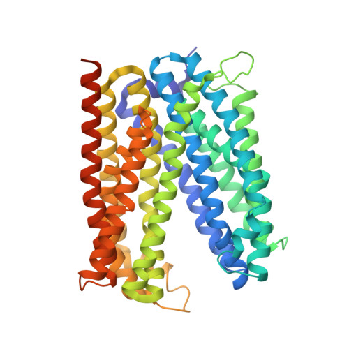
Inward-facing conformation of a multidrug resistance MATE family transporter.

Zakrzewska, S.Mehdipour, A.R.Malviya, V.N.Nonaka, T.Koepke, J.Muenke, C.Hausner, W.Hummer, G.Safarian, S.Michel, H.

(2019) Proc Natl Acad Sci U S A 116: 12275-12284

  • DOI: https://doi.org/10.1073/pnas.1904210116
  • Primary Citation Related Structures: 
    4MLB, 6FHZ, 6GWH, 6HFB

  • PubMed Abstract: 

    Multidrug and toxic compound extrusion (MATE) transporters mediate excretion of xenobiotics and toxic metabolites, thereby conferring multidrug resistance in bacterial pathogens and cancer cells. Structural information on the alternate conformational states and knowledge of the detailed mechanism of MATE transport are of great importance for drug development. However, the structures of MATE transporters are only known in V-shaped outward-facing conformations. Here, we present the crystal structure of a MATE transporter from Pyrococcus furiosus (PfMATE) in the long-sought-after inward-facing state, which was obtained after crystallization in the presence of native lipids. Transition from the outward-facing state to the inward-facing state involves rigid body movements of transmembrane helices (TMs) 2-6 and 8-12 to form an inverted V, facilitated by a loose binding of TM1 and TM7 to their respective bundles and their conformational flexibility. The inward-facing structure of PfMATE in combination with the outward-facing one supports an alternating access mechanism for the MATE family transporters.


  • Organizational Affiliation
    • Department of Molecular Membrane Biology, Max Planck Institute of Biophysics, 60438 Frankfurt am Main, Germany.

Macromolecule Content 

  • Total Structure Weight: 210.62 kDa 
  • Atom Count: 13,638 
  • Modeled Residue Count: 1,808 
  • Deposited Residue Count: 1,940 
  • Unique protein chains: 1

Macromolecules

Find similar proteins by:|  3D Structure
Entity ID: 1
MoleculeChains  Sequence LengthOrganismDetailsImage
Uncharacterized protein
A, B, C, D
485Pyrococcus furiosus DSM 3638Mutation(s): 1 
Gene Names: PF0708
Membrane Entity: Yes 
UniProt
Find proteins for Q8U2X0 (Pyrococcus furiosus (strain ATCC 43587 / DSM 3638 / JCM 8422 / Vc1))
Explore Q8U2X0 
Go to UniProtKB:  Q8U2X0
Entity Groups
Sequence Clusters30% Identity50% Identity70% Identity90% Identity95% Identity100% Identity
UniProt GroupQ8U2X0
Sequence Annotations
Expand
Reference Sequence

Small Molecules

Ligands 1 Unique
IDChains Name / Formula / InChI Key2D Diagram3D Interactions
CS

Query on CS


Download:Ideal Coordinates CCD File
E [auth A]
F [auth A]
G [auth A]
H [auth A]
I [auth B]
E [auth A],
F [auth A],
G [auth A],
H [auth A],
I [auth B],
J [auth B],
K [auth C],
L [auth C],
M [auth C],
N [auth D],
O [auth D],
P [auth D]
CESIUM ION
Cs
NCMHKCKGHRPLCM-UHFFFAOYSA-N

Experimental Data & Validation

Experimental Data

  • Method: X-RAY DIFFRACTION
  • Resolution: 3.50 Å
  • R-Value Free:  0.239 (Depositor), 0.245 (DCC) 
  • R-Value Work:  0.189 (Depositor), 0.201 (DCC) 
  • R-Value Observed: 0.192 (Depositor) 
Space Group: C 1 2 1
Unit Cell:
Length ( Å )Angle ( ˚ )
a = 219.752α = 90
b = 94.246β = 126.48
c = 138.785γ = 90
Software Package:
Software NamePurpose
PHENIXrefinement
XDSdata reduction
XSCALEdata scaling
PHASERphasing

Structure Validation

View Full Validation Report


Entry History 

& Funding Information

Deposition Data


Funding OrganizationLocationGrant Number
German Research FoundationGermany--
Max Planck SocietyGermany--

Revision History  (Full details and data files)

  • Version 1.0: 2019-06-12
    Type: Initial release
  • Version 1.1: 2019-06-26
    Changes: Data collection, Database references
  • Version 1.2: 2024-01-17
    Changes: Data collection, Database references, Derived calculations, Refinement description