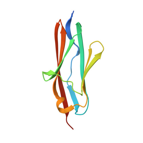
6HCI

Crystal structure of titin M3 domain


Experimental Data Snapshot

  • Method: X-RAY DIFFRACTION
  • Resolution: 2.12 Å
  • R-Value Free: 0.245 
  • R-Value Work: 0.192 
  • R-Value Observed: 0.194 

Starting Model: other
View more details

wwPDB Validation   3D Report Full Report


This is version 1.2 of the entry. See complete history


Literature

Structural diversity in the atomic resolution 3D fingerprint of the titin M-band segment.

Chatziefthimiou, S.D.Hornburg, P.Sauer, F.Mueller, S.Ugurlar, D.Xu, E.R.Wilmanns, M.

(2019) PLoS One 14: e0226693-e0226693

  • DOI: https://doi.org/10.1371/journal.pone.0226693
  • Primary Citation of Related Structures:  
    6H4L, 6HCI

  • PubMed Abstract: 

    In striated muscles, molecular filaments are largely composed of long protein chains with extensive arrays of identically folded domains, referred to as "beads-on-a-string". It remains a largely unresolved question how these domains have developed a unique molecular profile such that each carries out a distinct function without false-positive readout. This study focuses on the M-band segment of the sarcomeric protein titin, which comprises ten identically folded immunoglobulin domains. Comparative analysis of high-resolution structures of six of these domains ‒ M1, M3, M4, M5, M7, and M10 ‒ reveals considerable structural diversity within three distinct loops and a non-conserved pattern of exposed cysteines. Our data allow to structurally interpreting distinct pathological readouts that result from titinopathy-associated variants. Our findings support general principles that could be used to identify individual structural/functional profiles of hundreds of identically folded protein domains within the sarcomere and other densely crowded cellular environments.


  • Organizational Affiliation

    European Molecular Biology Laboratory, Hamburg Unit, Hamburg, Germany.


Macromolecules
Find similar proteins by:  (by identity cutoff)  |  3D Structure
Entity ID: 1
MoleculeChains Sequence LengthOrganismDetailsImage
Titin
A, B, C, D
105Homo sapiensMutation(s): 0 
Gene Names: TTN
EC: 2.7.11.1
UniProt & NIH Common Fund Data Resources
Find proteins for Q8WZ42 (Homo sapiens)
Explore Q8WZ42 
Go to UniProtKB:  Q8WZ42
PHAROS:  Q8WZ42
GTEx:  ENSG00000155657 
Entity Groups  
Sequence Clusters30% Identity50% Identity70% Identity90% Identity95% Identity100% Identity
UniProt GroupQ8WZ42
Sequence Annotations
Expand
  • Reference Sequence
Experimental Data & Validation

Experimental Data

  • Method: X-RAY DIFFRACTION
  • Resolution: 2.12 Å
  • R-Value Free: 0.245 
  • R-Value Work: 0.192 
  • R-Value Observed: 0.194 
  • Space Group: P 32 2 1
Unit Cell:
Length ( Å )Angle ( ˚ )
a = 106.061α = 90
b = 106.061β = 90
c = 75.481γ = 120
Software Package:
Software NamePurpose
PHENIXrefinement
XDSdata reduction
Aimlessdata scaling
PHASERphasing

Structure Validation

View Full Validation Report


Entry History 

Deposition Data

Revision History  (Full details and data files)

  • Version 1.0: 2019-08-28
    Type: Initial release
  • Version 1.1: 2020-01-01
    Changes: Database references
  • Version 1.2: 2024-05-01
    Changes: Data collection, Database references, Refinement description