6HCB

STRUCTURE OF GLUA2 LIGAND-BINDING DOMAIN (S1S2J-N775S) IN COMPLEX WITH GLUTAMATE AND TDPAM01 AT 1.9 A RESOLUTION.


Experimental Data Snapshot

  • Method: X-RAY DIFFRACTION
  • Resolution: 1.90 Å
  • R-Value Free: 0.192 
  • R-Value Work: 0.169 
  • R-Value Observed: 0.170 

wwPDB Validation   3D Report Full Report


Ligand Structure Quality Assessment 


This is version 1.2 of the entry. See complete history


Literature

Crystal Structures of Potent Dimeric Positive Allosteric Modulators at the Ligand-Binding Domain of the GluA2 Receptor.

Laulumaa, S.Hansen, K.V.Masternak, M.Drapier, T.Francotte, P.Pirotte, B.Frydenvang, K.Kastrup, J.S.

(2019) ACS Med Chem Lett 10: 243-247

  • DOI: https://doi.org/10.1021/acsmedchemlett.8b00369
  • Primary Citation of Related Structures:  
    6HC9, 6HCA, 6HCB, 6HCC, 6HCH

  • PubMed Abstract: 

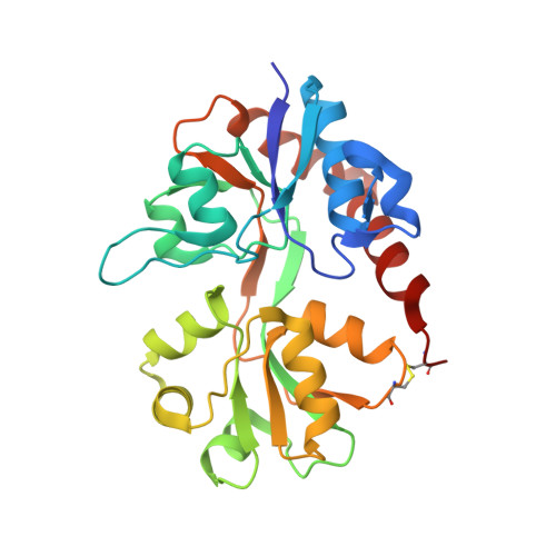
    The ionotropic glutamate receptor GluA2 is considered to be an attractive target for positive allosteric modulation for the development of pharmacological tools or cognitive enhancers. Here, we report a detailed structural characterization of two recently reported dimeric positive allosteric modulators, TDPAM01 and TDPAM02, with nanomolar potency at GluA2. Using X-ray crystallography, TDPAM01 and TDPAM02 were crystallized in the ligand-binding domain of the GluA2 flop isoform as well as in the flip-like mutant N775S and the preformed dimer L504Y-N775S. In all structures, one modulator molecule binds at the dimer interface with two characteristic hydrogen bonds being formed from the modulator to Pro515. Whereas the GluA2 dimers and modulator binding mode are similar when crystallized in the presence of l-glutamate, the shape of the binding site differs when no l-glutamate is present. TDPAM02 has no effect on domain closure in both apo and l-glutamate bound GluA2 dimers compared to structures without modulator.


  • Organizational Affiliation

    Department of Drug Design and Pharmacology, Faculty of Health and Medical Sciences, University of Copenhagen, Jagtvej 162, DK-2100 Copenhagen, Denmark.


Macromolecules
Find similar proteins by:  (by identity cutoff)  |  3D Structure
Entity ID: 1
MoleculeChains Sequence LengthOrganismDetailsImage
Glutamate receptor 2
A, B
264Rattus norvegicusMutation(s): 1 
Gene Names: Gria2Glur2
UniProt
Find proteins for P19491 (Rattus norvegicus)
Explore P19491 
Go to UniProtKB:  P19491
Entity Groups  
Sequence Clusters30% Identity50% Identity70% Identity90% Identity95% Identity100% Identity
UniProt GroupP19491
Sequence Annotations
Expand
  • Reference Sequence
Small Molecules
Ligands 6 Unique
IDChains Name / Formula / InChI Key2D Diagram3D Interactions
D45 (Subject of Investigation/LOI)
Query on D45

Download Ideal Coordinates CCD File 
S [auth B]6,6'-(Ethane-1,2-diyl)bis(4-methyl-3,4-dihydro-2H-1,2,4-benzothiadiazine 1,1-dioxide)
C18 H22 N4 O4 S2
PZUOXMWBXQXKJK-UHFFFAOYSA-N
GLU
Query on GLU

Download Ideal Coordinates CCD File 
I [auth A],
T [auth B]
GLUTAMIC ACID
C5 H9 N O4
WHUUTDBJXJRKMK-VKHMYHEASA-N
PEG
Query on PEG

Download Ideal Coordinates CCD File 
O [auth A],
X [auth B]
DI(HYDROXYETHYL)ETHER
C4 H10 O3
MTHSVFCYNBDYFN-UHFFFAOYSA-N
SO4
Query on SO4

Download Ideal Coordinates CCD File 
C [auth A]
D [auth A]
E [auth A]
F [auth A]
G [auth A]
C [auth A],
D [auth A],
E [auth A],
F [auth A],
G [auth A],
H [auth A],
P [auth B],
Q [auth B],
R [auth B]
SULFATE ION
O4 S
QAOWNCQODCNURD-UHFFFAOYSA-L
GOL
Query on GOL

Download Ideal Coordinates CCD File 
J [auth A],
K [auth A],
U [auth B],
V [auth B]
GLYCEROL
C3 H8 O3
PEDCQBHIVMGVHV-UHFFFAOYSA-N
CL
Query on CL

Download Ideal Coordinates CCD File 
L [auth A],
M [auth A],
N [auth A],
W [auth B]
CHLORIDE ION
Cl
VEXZGXHMUGYJMC-UHFFFAOYSA-M
Binding Affinity Annotations 
IDSourceBinding Affinity
D45 BindingDB:  6HCB EC50: min: 13, max: 14 (nM) from 2 assay(s)
Experimental Data & Validation

Experimental Data

  • Method: X-RAY DIFFRACTION
  • Resolution: 1.90 Å
  • R-Value Free: 0.192 
  • R-Value Work: 0.169 
  • R-Value Observed: 0.170 
  • Space Group: P 21 21 2
Unit Cell:
Length ( Å )Angle ( ˚ )
a = 97.436α = 90
b = 122.025β = 90
c = 47.623γ = 90
Software Package:
Software NamePurpose
PHENIXrefinement
SCALAdata scaling
PDB_EXTRACTdata extraction
XDSdata reduction
PHASERphasing

Structure Validation

View Full Validation Report


Ligand Structure Quality Assessment 


Entry History 

Deposition Data

Revision History  (Full details and data files)

  • Version 1.0: 2019-04-03
    Type: Initial release
  • Version 1.1: 2019-06-12
    Changes: Data collection, Structure summary
  • Version 1.2: 2024-01-17
    Changes: Data collection, Database references, Refinement description