ArabidopsisandChlamydomonasphosphoribulokinase crystal structures complete the redox structural proteome of the Calvin-Benson cycle.
Gurrieri, L., Del Giudice, A., Demitri, N., Falini, G., Pavel, N.V., Zaffagnini, M., Polentarutti, M., Crozet, P., Marchand, C.H., Henri, J., Trost, P., Lemaire, S.D., Sparla, F., Fermani, S.(2019) Proc Natl Acad Sci U S A 116: 8048-8053
- PubMed: 30923119 Search on PubMedSearch on PubMed Central
- DOI: https://doi.org/10.1073/pnas.1820639116
- Primary Citation Related Structures:
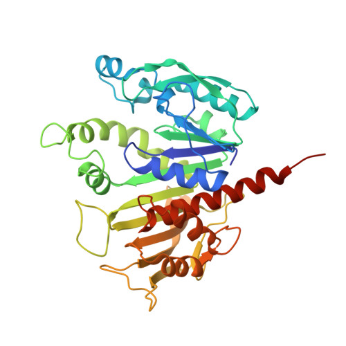
6H7G, 6H7H - PubMed Abstract:
In land plants and algae, the Calvin-Benson (CB) cycle takes place in the chloroplast, a specialized organelle in which photosynthesis occurs. Thioredoxins (TRXs) are small ubiquitous proteins, known to harmonize the two stages of photosynthesis through a thiol-based mechanism. Among the 11 enzymes of the CB cycle, the TRX target phosphoribulokinase (PRK) has yet to be characterized at the atomic scale. To accomplish this goal, we determined the crystal structures of PRK from two model species: the green alga Chlamydomonas reinhardtii ( Cr PRK) and the land plant Arabidopsis thaliana ( At PRK). PRK is an elongated homodimer characterized by a large central β-sheet of 18 strands, extending between two catalytic sites positioned at its edges. The electrostatic surface potential of the catalytic cavity has both a positive region suitable for binding the phosphate groups of substrates and an exposed negative region to attract positively charged TRX-f. In the catalytic cavity, the regulatory cysteines are 13 Å apart and connected by a flexible region exclusive to photosynthetic eukaryotes-the clamp loop-which is believed to be essential for oxidation-induced structural rearrangements. Structural comparisons with prokaryotic and evolutionarily older PRKs revealed that both At PRK and Cr PRK have a strongly reduced dimer interface and an increased number of random-coiled regions, suggesting that a general loss in structural rigidity correlates with gains in TRX sensitivity during the molecular evolution of PRKs in eukaryotes.
- Department of Pharmacy and Biotechnology-FaBiT, University of Bologna, 40126 Bologna, Italy.
Organizational Affiliation: