6H7B

Structure of Leishmania PABP1 (domain J) complexed with a peptide containing the PAM2 motif of eIF4E4.


Experimental Data Snapshot

  • Method: X-RAY DIFFRACTION
  • Resolution: 1.89 Å
  • R-Value Free: 0.230 
  • R-Value Work: 0.197 
  • R-Value Observed: 0.199 

wwPDB Validation   3D Report Full Report


This is version 1.2 of the entry. See complete history


Literature

The Leishmania PABP1-eIF4E4 interface: a novel 5'-3' interaction architecture for trans-spliced mRNAs.

Dos Santos Rodrigues, F.H.Firczuk, H.Breeze, A.L.Cameron, A.D.Walko, M.Wilson, A.J.Zanchin, N.I.T.McCarthy, J.E.G.

(2019) Nucleic Acids Res 47: 1493-1504

  • DOI: https://doi.org/10.1093/nar/gky1187
  • Primary Citation of Related Structures:  
    6H7A, 6H7B

  • PubMed Abstract: 

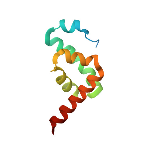
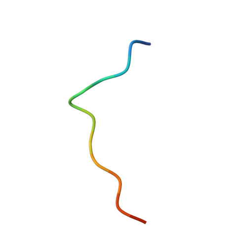
    Trans-splicing of trypanosomatid polycistronic transcripts produces polyadenylated monocistronic mRNAs modified to form the 5' cap4 structure (m7Gpppm36,6,2'Apm2'Apm2'Cpm23,2'U). NMR and X-ray crystallography reveal that Leishmania has a unique type of N-terminally-extended cap-binding protein (eIF4E4) that binds via a PAM2 motif to PABP1. This relies on the interactions of a combination of polar and charged amino acid side-chains together with multiple hydrophobic interactions, and underpins a novel architecture in the Leishmania cap4-binding translation factor complex. Measurements using microscale thermophoresis, fluorescence anisotropy and surface plasmon resonance characterize the key interactions driving assembly of the Leishmania translation initiation complex. We demonstrate that this complex can accommodate Leishmania eIF4G3 which, unlike the standard eukaryotic initiation complex paradigm, binds tightly to eIF4E4, but not to PABP1. Thus, in Leishmania, the chain of interactions 5'cap4-eIF4E4-PABP1-poly(A) bridges the mRNA 5' and 3' ends. Exceptionally, therefore, by binding tightly to two protein ligands and to the mRNA 5' cap4 structure, the trypanosomatid N-terminally extended form of eIF4E acts as the core molecular scaffold for the mRNA-cap-binding complex. Finally, the eIF4E4 N-terminal extension is an intrinsically disordered region that transitions to a partly folded form upon binding to PABP1, whereby this interaction is not modulated by poly(A) binding to PABP1.


  • Organizational Affiliation

    Warwick Integrative Synthetic Biology Centre (WISB) and School of Life Sciences, University of Warwick, Gibbet Hill, Coventry CV4 7AL, UK.


Macromolecules
Find similar proteins by:  (by identity cutoff)  |  3D Structure
Entity ID: 1
MoleculeChains Sequence LengthOrganismDetailsImage
Polyadenylate-binding protein
A, C
92Leishmania majorMutation(s): 0 
Gene Names: PABP1LMJF_35_5040
UniProt
Find proteins for E9AFX7 (Leishmania major)
Explore E9AFX7 
Go to UniProtKB:  E9AFX7
Entity Groups  
Sequence Clusters30% Identity50% Identity70% Identity90% Identity95% Identity100% Identity
UniProt GroupE9AFX7
Sequence Annotations
Expand
  • Reference Sequence

Find similar proteins by:  Sequence   |   3D Structure  

Entity ID: 2
MoleculeChains Sequence LengthOrganismDetailsImage
HIS-HIS-MET-ASN-PRO-ASN-ALA-THR-GLU-PHE-MET-PRO
B, D
14Leishmania majorMutation(s): 0 
UniProt
Find proteins for Q4Q7R3 (Leishmania major)
Explore Q4Q7R3 
Go to UniProtKB:  Q4Q7R3
Entity Groups  
Sequence Clusters30% Identity50% Identity70% Identity90% Identity95% Identity100% Identity
UniProt GroupQ4Q7R3
Sequence Annotations
Expand
  • Reference Sequence
Experimental Data & Validation

Experimental Data

  • Method: X-RAY DIFFRACTION
  • Resolution: 1.89 Å
  • R-Value Free: 0.230 
  • R-Value Work: 0.197 
  • R-Value Observed: 0.199 
  • Space Group: P 65
Unit Cell:
Length ( Å )Angle ( ˚ )
a = 83.33α = 90
b = 83.33β = 90
c = 58.16γ = 120
Software Package:
Software NamePurpose
PHENIXrefinement
XDSdata reduction
XSCALEdata scaling
PHASERphasing

Structure Validation

View Full Validation Report


Entry History & Funding Information

Deposition Data


Funding OrganizationLocationGrant Number
Medical Research Council (United Kingdom)United KingdomMR/N017447/1

Revision History  (Full details and data files)

  • Version 1.0: 2018-12-12
    Type: Initial release
  • Version 1.1: 2019-07-03
    Changes: Data collection, Database references
  • Version 1.2: 2024-01-17
    Changes: Data collection, Database references, Refinement description