6H45

crystal structure of the human TGT catalytic subunit QTRT1 in complex with queuine


Experimental Data Snapshot

  • Method: X-RAY DIFFRACTION
  • Resolution: 2.40 Å
  • R-Value Free: 0.240 
  • R-Value Work: 0.201 
  • R-Value Observed: 0.203 

wwPDB Validation   3D Report Full Report


Ligand Structure Quality Assessment 


This is version 2.0 of the entry. See complete history


Literature

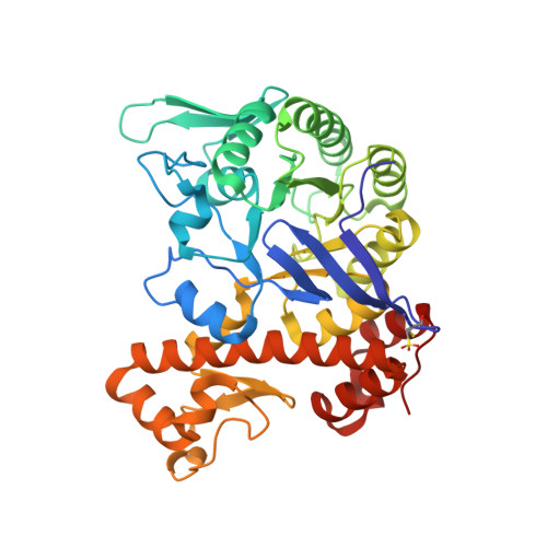
Crystal Structure of the Human tRNA Guanine Transglycosylase Catalytic Subunit QTRT1.

Johannsson, S.Neumann, P.Ficner, R.

(2018) Biomolecules 8

  • DOI: https://doi.org/10.3390/biom8030081
  • Primary Citation of Related Structures:  
    6H42, 6H45

  • PubMed Abstract: 

    RNA modifications have been implicated in diverse and important roles in all kingdoms of life with over 100 of them present on tRNAs. A prominent modification at the wobble base of four tRNAs is the 7-deaza-guanine derivative queuine which substitutes the guanine at position 34. This exchange is catalyzed by members of the enzyme class of tRNA guanine transglycosylases (TGTs). These enzymes incorporate guanine substituents into tRNA Asp , tRNA Asn tRNA His , and tRNA Tyr in all kingdoms of life. In contrast to the homodimeric bacterial TGT, the active eukaryotic TGT is a heterodimer in solution, comprised of a catalytic QTRT1 subunit and a noncatalytic QTRT2 subunit. Bacterial TGT enzymes, that incorporate a queuine precursor, have been identified or proposed as virulence factors for infections by pathogens in humans and therefore are valuable targets for drug design. To date no structure of a eukaryotic catalytic subunit is reported, and differences to its bacterial counterpart have to be deducted from sequence analysis and models. Here we report the first crystal structure of a eukaryotic QTRT1 subunit and compare it to known structures of the bacterial TGT and murine QTRT2. Furthermore, we were able to determine the crystal structure of QTRT1 in complex with the queuine substrate.


  • Organizational Affiliation

    Department of Molecular Structural Biology, Institute of Microbiology and Genetics, GZMB, Georg-August-University Göttingen, 37077 Göttingen, Germany. sjohann1@gwdg.de.


Macromolecules
Find similar proteins by:  (by identity cutoff)  |  3D Structure
Entity ID: 1
MoleculeChains Sequence LengthOrganismDetailsImage
Queuine tRNA-ribosyltransferase catalytic subunit 1
A, B
392Homo sapiensMutation(s): 0 
Gene Names: QTRT1TGTTGUT
EC: 2.4.2.64
UniProt & NIH Common Fund Data Resources
Find proteins for Q9BXR0 (Homo sapiens)
Explore Q9BXR0 
Go to UniProtKB:  Q9BXR0
PHAROS:  Q9BXR0
GTEx:  ENSG00000213339 
Entity Groups  
Sequence Clusters30% Identity50% Identity70% Identity90% Identity95% Identity100% Identity
UniProt GroupQ9BXR0
Sequence Annotations
Expand
  • Reference Sequence
Small Molecules
Ligands 6 Unique
IDChains Name / Formula / InChI Key2D Diagram3D Interactions
QEI
Query on QEI

Download Ideal Coordinates CCD File 
C [auth A],
K [auth B]
2-amino-5-({[(1S,4S,5R)-4,5-dihydroxycyclopent-2-en-1-yl]amino}methyl)-3,7-dihydro-4H-pyrrolo[2,3-d]pyrimidin-4-one
C12 H15 N5 O3
WYROLENTHWJFLR-ACLDMZEESA-N
GLU
Query on GLU

Download Ideal Coordinates CCD File 
N [auth B]GLUTAMIC ACID
C5 H9 N O4
WHUUTDBJXJRKMK-VKHMYHEASA-N
BR
Query on BR

Download Ideal Coordinates CCD File 
E [auth A],
I [auth A],
M [auth B],
R [auth B]
BROMIDE ION
Br
CPELXLSAUQHCOX-UHFFFAOYSA-M
ZN
Query on ZN

Download Ideal Coordinates CCD File 
D [auth A],
L [auth B]
ZINC ION
Zn
PTFCDOFLOPIGGS-UHFFFAOYSA-N
K
Query on K

Download Ideal Coordinates CCD File 
H [auth A],
Q [auth B]
POTASSIUM ION
K
NPYPAHLBTDXSSS-UHFFFAOYSA-N
CL
Query on CL

Download Ideal Coordinates CCD File 
F [auth A],
G [auth A],
J [auth B],
O [auth B],
P [auth B]
CHLORIDE ION
Cl
VEXZGXHMUGYJMC-UHFFFAOYSA-M
Modified Residues  1 Unique
IDChains TypeFormula2D DiagramParent
OCS
Query on OCS
A, B
L-PEPTIDE LINKINGC3 H7 N O5 SCYS
Experimental Data & Validation

Experimental Data

  • Method: X-RAY DIFFRACTION
  • Resolution: 2.40 Å
  • R-Value Free: 0.240 
  • R-Value Work: 0.201 
  • R-Value Observed: 0.203 
  • Space Group: C 1 2 1
Unit Cell:
Length ( Å )Angle ( ˚ )
a = 127.5α = 90
b = 158.86β = 108.19
c = 47.62γ = 90
Software Package:
Software NamePurpose
PHENIXrefinement
XDSdata reduction
XSCALEdata scaling
PHASERphasing
PDB_EXTRACTdata extraction

Structure Validation

View Full Validation Report


Ligand Structure Quality Assessment 


Entry History & Funding Information

Deposition Data


Funding OrganizationLocationGrant Number
German Research FoundationGermanySPP 1784

Revision History  (Full details and data files)

  • Version 1.0: 2018-09-05
    Type: Initial release
  • Version 2.0: 2023-03-15
    Changes: Advisory, Atomic model, Data collection, Database references, Derived calculations, Non-polymer description, Source and taxonomy, Structure summary