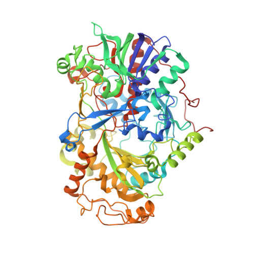
6H3O

Alcohol oxidase from Phanerochaete chrysosporium mutant F101S


Experimental Data Snapshot

  • Method: X-RAY DIFFRACTION
  • Resolution: 2.50 Å
  • R-Value Free: 0.286 
  • R-Value Work: 0.210 
  • R-Value Observed: 0.214 

Starting Model: experimental
View more details

wwPDB Validation   3D Report Full Report


Ligand Structure Quality Assessment 


This is version 1.3 of the entry. See complete history


Literature

Structure-Based Engineering of Phanerochaete chrysosporium Alcohol Oxidase for Enhanced Oxidative Power toward Glycerol.

Nguyen, Q.T.Romero, E.Dijkman, W.P.de Vasconcellos, S.P.Binda, C.Mattevi, A.Fraaije, M.W.

(2018) Biochemistry 57: 6209-6218

  • DOI: https://doi.org/10.1021/acs.biochem.8b00918
  • Primary Citation of Related Structures:  
    6H3G, 6H3O

  • PubMed Abstract: 

    Glycerol is a major byproduct of biodiesel production, and enzymes that oxidize this compound have been long sought after. The recently described alcohol oxidase from the white-rot basidiomycete Phanerochaete chrysosporium (PcAOX) was reported to feature very mild activity on glycerol. Here, we describe the comprehensive structural and biochemical characterization of this enzyme. PcAOX was expressed in Escherichia coli in high yields and displayed high thermostability. Steady-state kinetics revealed that PcAOX is highly active toward methanol, ethanol, and 1-propanol ( k cat = 18, 19, and 11 s -1 , respectively), but showed very limited activity toward glycerol ( k obs = 0.2 s -1 at 2 M substrate). The crystal structure of the homo-octameric PcAOX was determined at a resolution of 2.6 Å. The catalytic center is a remarkable solvent-inaccessible cavity located at the re side of the flavin cofactor. Its small size explains the observed preference for methanol and ethanol as best substrates. These findings led us to design several cavity-enlarging mutants with significantly improved activity toward glycerol. Among them, the F101S variant had a high k cat value of 3 s -1 , retaining a high degree of thermostability. The crystal structure of F101S PcAOX was solved, confirming the site of mutation and the larger substrate-binding pocket. Our data demonstrate that PcAOX is a very promising enzyme for glycerol biotransformation.


  • Organizational Affiliation

    Molecular Enzymology Group, Groningen Biomolecular Sciences and Biotechnology Institute , University of Groningen , Nijenborgh 4 , 9747 AG Groningen , The Netherlands.


Macromolecules
Find similar proteins by:  (by identity cutoff)  |  3D Structure
Entity ID: 1
MoleculeChains Sequence LengthOrganismDetailsImage
Alcohol oxidase
A, B, C, D, E
A, B, C, D, E, F, G, H
651Phanerodontia chrysosporiumMutation(s): 1 
Gene Names: aox1
UniProt
Find proteins for T2M2J4 (Phanerodontia chrysosporium)
Explore T2M2J4 
Go to UniProtKB:  T2M2J4
Entity Groups  
Sequence Clusters30% Identity50% Identity70% Identity90% Identity95% Identity100% Identity
UniProt GroupT2M2J4
Sequence Annotations
Expand
  • Reference Sequence
Small Molecules
Ligands 2 Unique
IDChains Name / Formula / InChI Key2D Diagram3D Interactions
FAD
Query on FAD

Download Ideal Coordinates CCD File 
I [auth A]
J [auth B]
L [auth C]
M [auth D]
N [auth E]
I [auth A],
J [auth B],
L [auth C],
M [auth D],
N [auth E],
O [auth F],
P [auth G],
Q [auth H]
FLAVIN-ADENINE DINUCLEOTIDE
C27 H33 N9 O15 P2
VWWQXMAJTJZDQX-UYBVJOGSSA-N
GOL
Query on GOL

Download Ideal Coordinates CCD File 
K [auth B],
R [auth H]
GLYCEROL
C3 H8 O3
PEDCQBHIVMGVHV-UHFFFAOYSA-N
Experimental Data & Validation

Experimental Data

Unit Cell:
Length ( Å )Angle ( ˚ )
a = 112.074α = 90
b = 203.553β = 104.64
c = 116.009γ = 90
Software Package:
Software NamePurpose
REFMACrefinement
XDSdata reduction
Aimlessdata scaling
MOLREPphasing

Structure Validation

View Full Validation Report


Ligand Structure Quality Assessment 


Entry History & Funding Information

Deposition Data


Funding OrganizationLocationGrant Number
European UnionItaly635734

Revision History  (Full details and data files)

  • Version 1.0: 2018-10-10
    Type: Initial release
  • Version 1.1: 2018-11-07
    Changes: Data collection, Database references, Structure summary
  • Version 1.2: 2020-04-22
    Changes: Database references
  • Version 1.3: 2024-01-17
    Changes: Data collection, Database references, Refinement description