6H1O

Structure of the BM3 heme domain in complex with voriconazole


Experimental Data Snapshot

  • Method: X-RAY DIFFRACTION
  • Resolution: 1.73 Å
  • R-Value Free: 0.200 
  • R-Value Work: 0.161 
  • R-Value Observed: 0.163 

wwPDB Validation   3D Report Full Report


Ligand Structure Quality Assessment 


This is version 1.1 of the entry. See complete history


Literature

Novel insights into P450 BM3 interactions with FDA-approved antifungal azole drugs.

Jeffreys, L.N.Poddar, H.Golovanova, M.Levy, C.W.Girvan, H.M.McLean, K.J.Voice, M.W.Leys, D.Munro, A.W.

(2019) Sci Rep 9: 1577-1577

  • DOI: https://doi.org/10.1038/s41598-018-37330-y
  • Primary Citation of Related Structures:  
    6H1L, 6H1O, 6H1S, 6H1T

  • PubMed Abstract: 

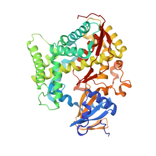
    Flavocytochrome P450 BM3 is a natural fusion protein constructed of cytochrome P450 and NADPH-cytochrome P450 reductase domains. P450 BM3 binds and oxidizes several mid- to long-chain fatty acids, typically hydroxylating these lipids at the ω-1, ω-2 and ω-3 positions. However, protein engineering has led to variants of this enzyme that are able to bind and oxidize diverse compounds, including steroids, terpenes and various human drugs. The wild-type P450 BM3 enzyme binds inefficiently to many azole antifungal drugs. However, we show that the BM3 A82F/F87V double mutant (DM) variant binds substantially tighter to numerous azole drugs than does the wild-type BM3, and that their binding occurs with more extensive heme spectral shifts indicative of complete binding of several azoles to the BM3 DM heme iron. We report here the first crystal structures of P450 BM3 bound to azole antifungal drugs - with the BM3 DM heme domain bound to the imidazole drugs clotrimazole and tioconazole, and to the triazole drugs fluconazole and voriconazole. This is the first report of any protein structure bound to the azole drug tioconazole, as well as the first example of voriconazole heme iron ligation through a pyrimidine nitrogen from its 5-fluoropyrimidine ring.


  • Organizational Affiliation

    Centre for Synthetic Biology of Fine and Specialty Chemicals (SYNBIOCHEM), Manchester Institute of Biotechnology, School of Chemistry, The University of Manchester, Manchester, M1 7DN, United Kingdom.


Macromolecules
Find similar proteins by:  (by identity cutoff)  |  3D Structure
Entity ID: 1
MoleculeChains Sequence LengthOrganismDetailsImage
Bifunctional cytochrome P450/NADPH--P450 reductase
A, B
457Priestia megateriumMutation(s): 2 
Gene Names: cyp102A1cyp102BG04_163
EC: 1.14.14.1 (PDB Primary Data), 1.6.2.4 (PDB Primary Data)
UniProt
Find proteins for P14779 (Priestia megaterium (strain ATCC 14581 / DSM 32 / CCUG 1817 / JCM 2506 / NBRC 15308 / NCIMB 9376 / NCTC 10342 / NRRL B-14308 / VKM B-512 / Ford 19))
Explore P14779 
Go to UniProtKB:  P14779
Entity Groups  
Sequence Clusters30% Identity50% Identity70% Identity90% Identity95% Identity100% Identity
UniProt GroupP14779
Sequence Annotations
Expand
  • Reference Sequence
Small Molecules
Ligands 5 Unique
IDChains Name / Formula / InChI Key2D Diagram3D Interactions
HEM
Query on HEM

Download Ideal Coordinates CCD File 
C [auth A],
L [auth B]
PROTOPORPHYRIN IX CONTAINING FE
C34 H32 Fe N4 O4
KABFMIBPWCXCRK-RGGAHWMASA-L
VOR (Subject of Investigation/LOI)
Query on VOR

Download Ideal Coordinates CCD File 
D [auth A],
M [auth B]
Voriconazole
C16 H14 F3 N5 O
BCEHBSKCWLPMDN-MGPLVRAMSA-N
PO4
Query on PO4

Download Ideal Coordinates CCD File 
E [auth A],
F [auth A],
G [auth A],
N [auth B],
O [auth B]
PHOSPHATE ION
O4 P
NBIIXXVUZAFLBC-UHFFFAOYSA-K
GOL
Query on GOL

Download Ideal Coordinates CCD File 
H [auth A],
I [auth A],
J [auth A],
P [auth B]
GLYCEROL
C3 H8 O3
PEDCQBHIVMGVHV-UHFFFAOYSA-N
EDO
Query on EDO

Download Ideal Coordinates CCD File 
K [auth A]1,2-ETHANEDIOL
C2 H6 O2
LYCAIKOWRPUZTN-UHFFFAOYSA-N
Experimental Data & Validation

Experimental Data

  • Method: X-RAY DIFFRACTION
  • Resolution: 1.73 Å
  • R-Value Free: 0.200 
  • R-Value Work: 0.161 
  • R-Value Observed: 0.163 
  • Space Group: P 1 21 1
Unit Cell:
Length ( Å )Angle ( ˚ )
a = 59.853α = 90
b = 150.009β = 96.85
c = 61.069γ = 90
Software Package:
Software NamePurpose
PHENIXrefinement
XDSdata reduction
XDSdata scaling
PHASERphasing

Structure Validation

View Full Validation Report


Ligand Structure Quality Assessment 


Entry History & Funding Information

Deposition Data


Funding OrganizationLocationGrant Number
Biotechnology and Biological Sciences Research CouncilUnited KingdomBB/K001884/1

Revision History  (Full details and data files)

  • Version 1.0: 2019-02-20
    Type: Initial release
  • Version 1.1: 2024-01-17
    Changes: Data collection, Database references, Derived calculations, Refinement description