6GX3 | pdb_00006gx3

Crystal structure of Schistosoma mansoni HDAC8 complexed with an hydroxamate 1


Experimental Data Snapshot

  • Method: X-RAY DIFFRACTION
  • Resolution: 2.10 Å
  • R-Value Free: 
    0.239 (Depositor), 0.225 (DCC) 
  • R-Value Work: 
    0.189 (Depositor) 
  • R-Value Observed: 
    0.192 (Depositor) 

Starting Model: experimental
View more details

wwPDB Validation 3D Report Full Report

Validation slider image for 6GX3

Ligand Structure Quality Assessment 


This is version 1.2 of the entry. See complete history

Literature

Synthesis, Crystallization Studies, and in vitro Characterization of Cinnamic Acid Derivatives as SmHDAC8 Inhibitors for the Treatment of Schistosomiasis.

Bayer, T.Chakrabarti, A.Lancelot, J.Shaik, T.B.Hausmann, K.Melesina, J.Schmidtkunz, K.Marek, M.Erdmann, F.Schmidt, M.Robaa, D.Romier, C.Pierce, R.J.Jung, M.Sippl, W.

(2018) ChemMedChem 13: 1517-1529

  • DOI: https://doi.org/10.1002/cmdc.201800238
  • Primary Citation Related Structures: 
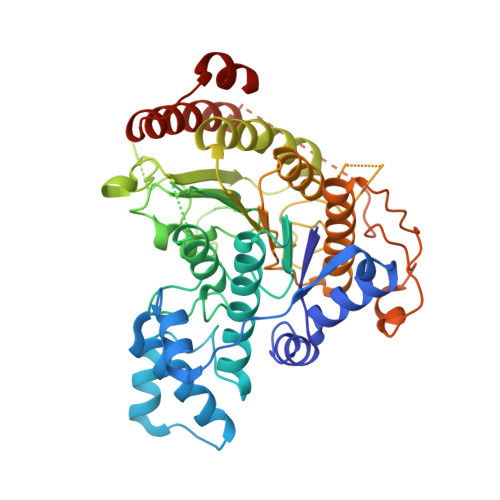
    6GX3, 6GXA, 6GXU, 6GXW

  • PubMed Abstract: 

    Schistosomiasis is a neglected parasitic disease that affects more than 265 million people worldwide and for which the control strategy relies on mass treatment with only one drug: praziquantel. Based on the 3-chlorobenzothiophene-2-hydroxamic acid J1075, a series of hydroxamic acids with different scaffolds were prepared as potential inhibitors of Schistosoma mansoni histone deacetylase 8 (SmHDAC8). The crystal structures of SmHDAC8 with four inhibitors provided insight into the binding mode and orientation of molecules in the binding pocket as well as the orientation of its flexible amino acid residues. The compounds were evaluated in screens for inhibitory activity against schistosome and human HDACs. The most promising compounds were further investigated for their activity toward the major human HDAC isotypes. The most potent inhibitors were additionally screened for lethality against the schistosome larval stage using a fluorescence-based assay. Two of the compounds showed significant, dose-dependent killing of the schistosome larvae and markedly impaired egg laying of adult worm pairs maintained in culture.


  • Organizational Affiliation
    • Institute of Pharmacy, Martin Luther University of Halle-Wittenberg, Wolfgang-Langenbeck-Str. 4, 06120, Halle/Saale, Germany.

Macromolecule Content 

  • Total Structure Weight: 205.43 kDa 
  • Atom Count: 13,570 
  • Modeled Residue Count: 1,612 
  • Deposited Residue Count: 1,788 
  • Unique protein chains: 1

Macromolecules

Find similar proteins by:|  3D Structure
Entity ID: 1
MoleculeChains  Sequence LengthOrganismDetailsImage
Histone deacetylase
A, B, C, D
447Schistosoma mansoniMutation(s): 0 
Gene Names: HDAC8
EC: 3.5.1.98
UniProt
Find proteins for A5H660 (Schistosoma mansoni)
Explore A5H660 
Go to UniProtKB:  A5H660
Entity Groups
Sequence Clusters30% Identity50% Identity70% Identity90% Identity95% Identity100% Identity
UniProt GroupA5H660
Sequence Annotations
Expand
Reference Sequence

Small Molecules

Ligands 5 Unique
IDChains Name / Formula / InChI Key2D Diagram3D Interactions
FF2
(Subject of Investigation/LOI)

Query on FF2


Download:Ideal Coordinates CCD File
EA [auth D]
FA [auth D]
H [auth A]
I [auth A]
O [auth B]
EA [auth D],
FA [auth D],
H [auth A],
I [auth A],
O [auth B],
P [auth B],
W [auth C],
X [auth C]
4-chloranyl-~{N}-oxidanyl-1-benzothiophene-2-carboxamide
C9 H6 Cl N O2 S
IHGVZTXLHUWVIA-UHFFFAOYSA-N
GOL

Query on GOL


Download:Ideal Coordinates CCD File
J [auth A]
K [auth A]
Q [auth B]
R [auth B]
Y [auth C]
J [auth A],
K [auth A],
Q [auth B],
R [auth B],
Y [auth C],
Z [auth C]
GLYCEROL
C3 H8 O3
PEDCQBHIVMGVHV-UHFFFAOYSA-N
DMF

Query on DMF


Download:Ideal Coordinates CCD File
AA [auth C],
S [auth B]
DIMETHYLFORMAMIDE
C3 H7 N O
ZMXDDKWLCZADIW-UHFFFAOYSA-N
ZN

Query on ZN


Download:Ideal Coordinates CCD File
BA [auth D],
E [auth A],
L [auth B],
T [auth C]
ZINC ION
Zn
PTFCDOFLOPIGGS-UHFFFAOYSA-N
K

Query on K


Download:Ideal Coordinates CCD File
CA [auth D]
DA [auth D]
F [auth A]
G [auth A]
M [auth B]
CA [auth D],
DA [auth D],
F [auth A],
G [auth A],
M [auth B],
N [auth B],
U [auth C],
V [auth C]
POTASSIUM ION
K
NPYPAHLBTDXSSS-UHFFFAOYSA-N

Experimental Data & Validation

Experimental Data

  • Method: X-RAY DIFFRACTION
  • Resolution: 2.10 Å
  • R-Value Free:  0.239 (Depositor), 0.225 (DCC) 
  • R-Value Work:  0.189 (Depositor) 
  • R-Value Observed: 0.192 (Depositor) 
Space Group: P 1
Unit Cell:
Length ( Å )Angle ( ˚ )
a = 70.449α = 75.71
b = 70.67β = 78.4
c = 98.413γ = 85.55
Software Package:
Software NamePurpose
PHENIXrefinement
XDSdata reduction
XDSdata scaling
PHENIXphasing

Structure Validation

View Full Validation Report


Ligand Structure Quality Assessment 


Entry History 

& Funding Information

Deposition Data


Funding OrganizationLocationGrant Number
European CommissionFrance241865
European CommissionFrance602080

Revision History  (Full details and data files)

  • Version 1.0: 2018-08-15
    Type: Initial release
  • Version 1.1: 2018-08-29
    Changes: Data collection, Database references
  • Version 1.2: 2024-01-17
    Changes: Data collection, Database references, Derived calculations, Refinement description