6GVC | pdb_00006gvc

Structure of ArhGAP12 bound to G-Actin


Experimental Data Snapshot

  • Method: X-RAY DIFFRACTION
  • Resolution: 2.60 Å
  • R-Value Free: 
    0.251 (Depositor), 0.252 (DCC) 
  • R-Value Work: 
    0.207 (Depositor), 0.209 (DCC) 
  • R-Value Observed: 
    0.209 (Depositor) 

Starting Model: experimental
View more details

wwPDB Validation 3D Report Full Report

Validation slider image for 6GVC

Ligand Structure Quality Assessment 


This is version 1.2 of the entry. See complete history

Literature

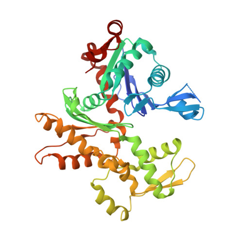
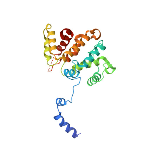
RPEL-family rhoGAPs link Rac/Cdc42 GTP loading to G-actin availability.

Diring, J.Mouilleron, S.McDonald, N.Q.Treisman, R.

(2019) Nat Cell Biol 21: 845-855

  • DOI: https://doi.org/10.1038/s41556-019-0337-y
  • Primary Citation Related Structures: 
    6GVC

  • PubMed Abstract: 

    RPEL proteins, which contain the G-actin-binding RPEL motif, coordinate cytoskeletal processes with actin dynamics. We show that the ArhGAP12- and ArhGAP32-family GTPase-activating proteins (GAPs) are RPEL proteins. We determine the structure of the ArhGAP12/G-actin complex, and show that G-actin contacts the RPEL motif and GAP domain sequences. G-actin inhibits ArhGAP12 GAP activity, and this requires the G-actin contacts identified in the structure. In B16 melanoma cells, ArhGAP12 suppresses basal Rac and Cdc42 activity, F-actin assembly, invadopodia formation and experimental metastasis. In this setting, ArhGAP12 mutants defective for G-actin binding exhibit more effective downregulation of Rac GTP loading following HGF stimulation and enhanced inhibition of Rac-dependent processes, including invadopodia formation. Potentiation or disruption of the G-actin/ArhGAP12 interaction, by treatment with the actin-binding drugs latrunculin B or cytochalasin D, has corresponding effects on Rac GTP loading. The interaction of G-actin with RPEL-family rhoGAPs thus provides a negative feedback loop that couples Rac activity to actin dynamics.


  • Organizational Affiliation
    • Signalling and Transcription Group, The Francis Crick Institute, London, UK.

Macromolecule Content 

  • Total Structure Weight: 279.27 kDa 
  • Atom Count: 18,354 
  • Modeled Residue Count: 2,308 
  • Deposited Residue Count: 2,432 
  • Unique protein chains: 2

Macromolecules

Find similar proteins by:|  3D Structure
Entity ID: 1
MoleculeChains  Sequence LengthOrganismDetailsImage
Actin, alpha skeletal muscleA [auth B],
B [auth A],
C,
D
377Oryctolagus cuniculusMutation(s): 0 
EC: 3.6.4
UniProt
Find proteins for P68135 (Oryctolagus cuniculus)
Explore P68135 
Go to UniProtKB:  P68135
Entity Groups
Sequence Clusters30% Identity50% Identity70% Identity90% Identity95% Identity100% Identity
UniProt GroupP68135
Sequence Annotations
Expand
Reference Sequence
Find similar proteins by:|  3D Structure
Entity ID: 2
MoleculeChains  Sequence LengthOrganismDetailsImage
Rho GTPase-activating protein 12E [auth R],
F [auth Q],
G [auth S],
H [auth T]
231Mus musculusMutation(s): 0 
Gene Names: Arhgap12
UniProt & NIH Common Fund Data Resources
Find proteins for Q8C0D4 (Mus musculus)
Explore Q8C0D4 
Go to UniProtKB:  Q8C0D4
IMPC:  MGI:1922665
Entity Groups
Sequence Clusters30% Identity50% Identity70% Identity90% Identity95% Identity100% Identity
UniProt GroupQ8C0D4
Sequence Annotations
Expand
Reference Sequence

Small Molecules

Ligands 4 Unique
IDChains Name / Formula / InChI Key2D Diagram3D Interactions
ATP

Query on ATP


Download:Ideal Coordinates CCD File
K [auth B],
O [auth A],
S [auth C],
W [auth D]
ADENOSINE-5'-TRIPHOSPHATE
C10 H16 N5 O13 P3
ZKHQWZAMYRWXGA-KQYNXXCUSA-N
LAB

Query on LAB


Download:Ideal Coordinates CCD File
J [auth B],
N [auth A],
R [auth C],
V [auth D]
LATRUNCULIN B
C20 H29 N O5 S
NSHPHXHGRHSMIK-JRIKCGFMSA-N
EDO

Query on EDO


Download:Ideal Coordinates CCD File
AA [auth S]
BA [auth S]
I [auth B]
M [auth A]
Q [auth C]
AA [auth S],
BA [auth S],
I [auth B],
M [auth A],
Q [auth C],
U [auth D],
Y [auth R],
Z [auth Q]
1,2-ETHANEDIOL
C2 H6 O2
LYCAIKOWRPUZTN-UHFFFAOYSA-N
MG

Query on MG


Download:Ideal Coordinates CCD File
L [auth B],
P [auth A],
T [auth C],
X [auth D]
MAGNESIUM ION
Mg
JLVVSXFLKOJNIY-UHFFFAOYSA-N

Experimental Data & Validation

Experimental Data

  • Method: X-RAY DIFFRACTION
  • Resolution: 2.60 Å
  • R-Value Free:  0.251 (Depositor), 0.252 (DCC) 
  • R-Value Work:  0.207 (Depositor), 0.209 (DCC) 
  • R-Value Observed: 0.209 (Depositor) 
Space Group: P 1 21 1
Unit Cell:
Length ( Å )Angle ( ˚ )
a = 101.65α = 90
b = 130.24β = 111.17
c = 109.3γ = 90
Software Package:
Software NamePurpose
PHENIXrefinement
DIALSdata reduction
Aimlessdata scaling
PHASERphasing

Structure Validation

View Full Validation Report


Ligand Structure Quality Assessment 


Entry History 

& Funding Information

Deposition Data


Funding OrganizationLocationGrant Number
European Research CouncilUnited Kingdom268690

Revision History  (Full details and data files)

  • Version 1.0: 2019-03-27
    Type: Initial release
  • Version 1.1: 2019-10-23
    Changes: Data collection, Database references
  • Version 1.2: 2024-01-17
    Changes: Data collection, Database references, Derived calculations, Refinement description