6GOW | pdb_00006gow

Crystal structure of the flagellin-FliS complex from Bacillus subtilis crystallized in spacegroup P22121


Experimental Data Snapshot

  • Method: X-RAY DIFFRACTION
  • Resolution: 2.10 Å
  • R-Value Free: 
    0.204 (Depositor), 0.203 (DCC) 
  • R-Value Work: 
    0.175 (Depositor), 0.176 (DCC) 
  • R-Value Observed: 
    0.177 (Depositor) 

Starting Model: experimental
View more details

wwPDB Validation 3D Report Full Report

Validation slider image for 6GOW

This is version 1.1 of the entry. See complete history

Literature

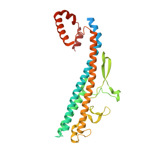
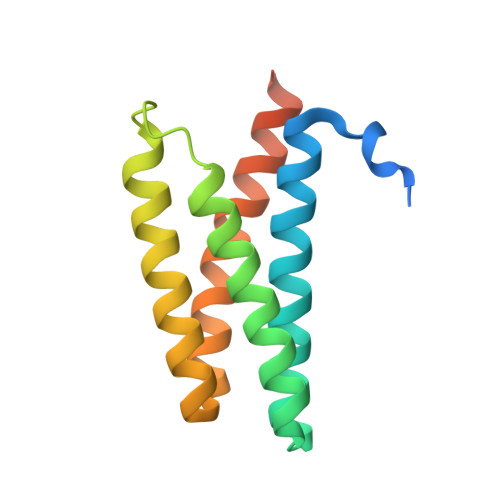
FliS/flagellin/FliW heterotrimer couples type III secretion and flagellin homeostasis.

Altegoer, F.Mukherjee, S.Steinchen, W.Bedrunka, P.Linne, U.Kearns, D.B.Bange, G.

(2018) Sci Rep 8: 11552-11552

  • DOI: https://doi.org/10.1038/s41598-018-29884-8
  • Primary Citation Related Structures: 
    5MAW, 6GOW

  • PubMed Abstract: 

    Flagellin is amongst the most abundant proteins in flagellated bacterial species and constitutes the major building block of the flagellar filament. The proteins FliW and FliS serve in the post-transcriptional control of flagellin and guide the protein to the flagellar type III secretion system (fT3SS), respectively. Here, we present the high-resolution structure of FliS/flagellin heterodimer and show that FliS and FliW bind to opposing interfaces located at the N- and C-termini of flagellin. The FliS/flagellin/FliW heterotrimer is able to interact with FlhA-C suggesting that FliW and FliS are released during flagellin export. After release, FliW and FliS are recycled to execute a new round of post-transcriptional regulation and targeting. Taken together, our study provides a mechanism explaining how FliW and FliS synchronize the production of flagellin with the capacity of the fT3SS to secrete flagellin.


  • Organizational Affiliation
    • LOEWE Center for Synthetic Microbiology & Dep. of Chemistry, Philipps University Marburg, Hans-Meerwein-Strasse 6, 35043, Marburg, Germany.

Macromolecule Content 

  • Total Structure Weight: 47.81 kDa 
  • Atom Count: 3,149 
  • Modeled Residue Count: 366 
  • Deposited Residue Count: 437 
  • Unique protein chains: 2

Macromolecules

Find similar proteins by:|  3D Structure
Entity ID: 1
MoleculeChains  Sequence LengthOrganismDetailsImage
FlagellinA [auth D]304Bacillus subtilisMutation(s): 0 
Gene Names: B4417_3365
UniProt
Find proteins for P02968 (Bacillus subtilis (strain 168))
Explore P02968 
Go to UniProtKB:  P02968
Entity Groups
Sequence Clusters30% Identity50% Identity70% Identity90% Identity95% Identity100% Identity
UniProt GroupP02968
Sequence Annotations
Expand
Reference Sequence
Find similar proteins by:|  3D Structure
Entity ID: 2
MoleculeChains  Sequence LengthOrganismDetailsImage
Flagellar secretion chaperone FliSB [auth E]133Bacillus subtilisMutation(s): 0 
Gene Names: B4417_3362SC09_contig4orf00739
UniProt
Find proteins for P39739 (Bacillus subtilis (strain 168))
Explore P39739 
Go to UniProtKB:  P39739
Entity Groups
Sequence Clusters30% Identity50% Identity70% Identity90% Identity95% Identity100% Identity
UniProt GroupP39739
Sequence Annotations
Expand
Reference Sequence

Experimental Data & Validation

Experimental Data

  • Method: X-RAY DIFFRACTION
  • Resolution: 2.10 Å
  • R-Value Free:  0.204 (Depositor), 0.203 (DCC) 
  • R-Value Work:  0.175 (Depositor), 0.176 (DCC) 
  • R-Value Observed: 0.177 (Depositor) 
Space Group: P 2 21 21
Unit Cell:
Length ( Å )Angle ( ˚ )
a = 50.86α = 90
b = 89.956β = 90
c = 116.079γ = 90
Software Package:
Software NamePurpose
PHENIXrefinement
XDSdata reduction
SCALAdata scaling
PHASERphasing

Structure Validation

View Full Validation Report


Entry History 

Deposition Data

Revision History  (Full details and data files)

  • Version 1.0: 2018-08-29
    Type: Initial release
  • Version 1.1: 2024-01-17
    Changes: Data collection, Database references, Refinement description