6GMA

Crystal structure of the FIP200 C-terminal region


Experimental Data Snapshot

  • Method: X-RAY DIFFRACTION
  • Resolution: 3.20 Å
  • R-Value Free: 0.295 
  • R-Value Work: 0.267 
  • R-Value Observed: 0.269 

wwPDB Validation   3D Report Full Report


This is version 1.2 of the entry. See complete history


Literature

FIP200 Claw Domain Binding to p62 Promotes Autophagosome Formation at Ubiquitin Condensates.

Turco, E.Witt, M.Abert, C.Bock-Bierbaum, T.Su, M.Y.Trapannone, R.Sztacho, M.Danieli, A.Shi, X.Zaffagnini, G.Gamper, A.Schuschnig, M.Fracchiolla, D.Bernklau, D.Romanov, J.Hartl, M.Hurley, J.H.Daumke, O.Martens, S.

(2019) Mol Cell 74: 330-346.e11

  • DOI: https://doi.org/10.1016/j.molcel.2019.01.035
  • Primary Citation of Related Structures:  
    6DCE, 6GMA

  • PubMed Abstract: 

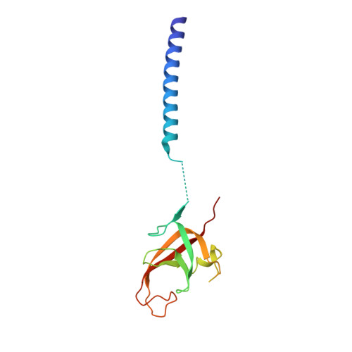
    The autophagy cargo receptor p62 facilitates the condensation of misfolded, ubiquitin-positive proteins and their degradation by autophagy, but the molecular mechanism of p62 signaling to the core autophagy machinery is unclear. Here, we show that disordered residues 326-380 of p62 directly interact with the C-terminal region (CTR) of FIP200. Crystal structure determination shows that the FIP200 CTR contains a dimeric globular domain that we designated the "Claw" for its shape. The interaction of p62 with FIP200 is mediated by a positively charged pocket in the Claw, enhanced by p62 phosphorylation, mutually exclusive with the binding of p62 to LC3B, and it promotes degradation of ubiquitinated cargo by autophagy. Furthermore, the recruitment of the FIP200 CTR slows the phase separation of ubiquitinated proteins by p62 in a reconstituted system. Our data provide the molecular basis for a crosstalk between cargo condensation and autophagosome formation.


  • Organizational Affiliation

    Department of Biochemistry and Cell Biology, Max F. Perutz Laboratories (MFPL), University of Vienna, Vienna BioCenter, Dr. Bohr-Gasse 9, 1030 Vienna, Austria.


Macromolecules
Find similar proteins by:  (by identity cutoff)  |  3D Structure
Entity ID: 1
MoleculeChains Sequence LengthOrganismDetailsImage
RB1-inducible coiled-coil protein 1
A, B, C, D, E
A, B, C, D, E, F
140Homo sapiensMutation(s): 0 
Gene Names: RB1CC1KIAA0203RBICC
UniProt & NIH Common Fund Data Resources
Find proteins for Q8TDY2 (Homo sapiens)
Explore Q8TDY2 
Go to UniProtKB:  Q8TDY2
PHAROS:  Q8TDY2
GTEx:  ENSG00000023287 
Entity Groups  
Sequence Clusters30% Identity50% Identity70% Identity90% Identity95% Identity100% Identity
UniProt GroupQ8TDY2
Sequence Annotations
Expand
  • Reference Sequence
Experimental Data & Validation

Experimental Data

  • Method: X-RAY DIFFRACTION
  • Resolution: 3.20 Å
  • R-Value Free: 0.295 
  • R-Value Work: 0.267 
  • R-Value Observed: 0.269 
  • Space Group: P 21 21 2
Unit Cell:
Length ( Å )Angle ( ˚ )
a = 92.045α = 90
b = 187.166β = 90
c = 55.343γ = 90
Software Package:
Software NamePurpose
PHENIXrefinement
XDSdata reduction
XSCALEdata scaling
PDB_EXTRACTdata extraction
AutoSolphasing

Structure Validation

View Full Validation Report


Entry History & Funding Information

Deposition Data


Funding OrganizationLocationGrant Number
European Research CouncilGermanyERC-2013-CoG-616024

Revision History  (Full details and data files)

  • Version 1.0: 2019-03-06
    Type: Initial release
  • Version 1.1: 2019-04-24
    Changes: Data collection, Database references
  • Version 1.2: 2019-05-01
    Changes: Data collection, Database references