Reaction of O2with a diiron protein generates a mixed-valent Fe2+/Fe3+center and peroxide.
Bradley, J.M., Svistunenko, D.A., Pullin, J., Hill, N., Stuart, R.K., Palenik, B., Wilson, M.T., Hemmings, A.M., Moore, G.R., Le Brun, N.E.(2019) Proc Natl Acad Sci U S A 116: 2058-2067
- PubMed: 30659147 Search on PubMedSearch on PubMed Central
- DOI: https://doi.org/10.1073/pnas.1809913116
- Primary Citation Related Structures:
5OUW, 5OUZ, 6GKA, 6GKB, 6GKC - PubMed Abstract:
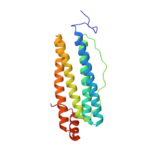
The gene encoding the cyanobacterial ferritin Syn Ftn is up-regulated in response to copper stress. Here, we show that, while Syn Ftn does not interact directly with copper, it is highly unusual in several ways. First, its catalytic diiron ferroxidase center is unlike those of all other characterized prokaryotic ferritins and instead resembles an animal H-chain ferritin center. Second, as demonstrated by kinetic, spectroscopic, and high-resolution X-ray crystallographic data, reaction of O 2 with the di-Fe 2+ center results in a direct, one-electron oxidation to a mixed-valent Fe 2+ /Fe 3+ form. Iron-O 2 chemistry of this type is currently unknown among the growing family of proteins that bind a diiron site within a four α-helical bundle in general and ferritins in particular. The mixed-valent form, which slowly oxidized to the more usual di-Fe 3+ form, is an intermediate that is continually generated during mineralization. Peroxide, rather than superoxide, is shown to be the product of O 2 reduction, implying that ferroxidase centers function in pairs via long-range electron transfer through the protein resulting in reduction of O 2 bound at only one of the centers. We show that electron transfer is mediated by the transient formation of a radical on Tyr40, which lies ∼4 Å from the diiron center. As well as demonstrating an expansion of the iron-O 2 chemistry known to occur in nature, these data are also highly relevant to the question of whether all ferritins mineralize iron via a common mechanism, providing unequivocal proof that they do not.
- Centre for Molecular and Structural Biochemistry, School of Chemistry, University of East Anglia, Norwich NR4 7TJ, United Kingdom.
Organizational Affiliation: