6GJT

Chlamydia protein Pgp3 studied at high resolution in a new crystal form


Experimental Data Snapshot

  • Method: X-RAY DIFFRACTION
  • Resolution: 1.98 Å
  • R-Value Free: 0.292 
  • R-Value Work: 0.273 
  • R-Value Observed: 0.274 

wwPDB Validation   3D Report Full Report


This is version 1.2 of the entry. See complete history


Literature

Chlamydia protein Pgp3 studied at high resolution in a new crystal form.

Khurshid, S.Govada, L.Wills, G.McClure, M.O.Helliwell, J.R.Chayen, N.E.

(2018) IUCrJ 5: 439-448

  • DOI: https://doi.org/10.1107/S2052252518007637
  • Primary Citation of Related Structures:  
    6GJT

  • PubMed Abstract: 

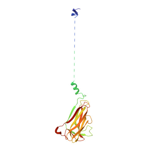
    The protein Pgp3 is implicated in the sexually transmitted disease chlamydia and comprises an extended complex arrangement of a C-terminal domain (CTD) and an N-terminal domain (NTD) linked by a triple-helix coiled coil (THCC). Here, the X-ray crystal structure of Pgp3 from an LGV1 strain is reported at the highest X-ray diffraction resolution obtained to date for the full protein. The protein was crystallized using a high concentration of potassium bromide, which resulted in a new crystal form with relatively low solvent content that diffracted to a resolution of 1.98 Å. The three-dimensional structure of this new crystal form is described and compared with those of other crystal forms, and the potassium bromide binding sites and the relevance to chlamydia isolates from around the globe are described. The crystal packing is apparently driven by the CTDs. Since the threefold axes of the THCC and NTD are not collinear with the threefold axis of a CTD, this naturally leads to disorder in the THCC and the portion of the NTD that does not directly interact with the CTD via crystal packing. The key avenue to resolving these oddities in the crystal structure analysis was a complete new analysis in space group P 1 and determining the space group as P 2 1 2 1 2 1 . This space-group assignment was that originally determined from the diffraction pattern but was perhaps complicated by translational noncrystallographic symmetry. This crystal structure of a three-domain multi-macromolecular complex with two misaligned threefold axes was a unique challenge and has not been encountered before. It is suggested that a specific intermolecular interaction, possibly of functional significance in receptor binding in chlamydia, might allow the design of a new chemotherapeutic agent against chlamydia.


  • Organizational Affiliation

    Computational and Systems Medicine, Department of Surgery and Cancer, Imperial College London, Sir Alexander Fleming Building, South Kensington, London SW7 2AZ, England.


Macromolecules
Find similar proteins by:  (by identity cutoff)  |  3D Structure
Entity ID: 1
MoleculeChains Sequence LengthOrganismDetailsImage
Virulence plasmid protein pGP3-D
A, B, C, D, E
A, B, C, D, E, F
264Chlamydia trachomatisMutation(s): 0 
UniProt
Find proteins for P0CE18 (Chlamydia trachomatis)
Explore P0CE18 
Go to UniProtKB:  P0CE18
Entity Groups  
Sequence Clusters30% Identity50% Identity70% Identity90% Identity95% Identity100% Identity
UniProt GroupP0CE18
Sequence Annotations
Expand
  • Reference Sequence
Small Molecules
Ligands 2 Unique
IDChains Name / Formula / InChI Key2D Diagram3D Interactions
BR
Query on BR

Download Ideal Coordinates CCD File 
CA [auth E]
DA [auth E]
EA [auth E]
FA [auth E]
G [auth A]
CA [auth E],
DA [auth E],
EA [auth E],
FA [auth E],
G [auth A],
GA [auth E],
H [auth A],
HA [auth E],
I [auth A],
IA [auth E],
JA [auth F],
KA [auth F],
LA [auth F],
M [auth B],
MA [auth F],
N [auth B],
O [auth B],
P [auth B],
Q [auth B],
R [auth B],
S [auth C],
T [auth C],
U [auth C],
V [auth C],
W [auth C],
X [auth D],
Y [auth D],
Z [auth D]
BROMIDE ION
Br
CPELXLSAUQHCOX-UHFFFAOYSA-M
K
Query on K

Download Ideal Coordinates CCD File 
AA [auth D]
BA [auth D]
J [auth A]
K [auth A]
L [auth A]
AA [auth D],
BA [auth D],
J [auth A],
K [auth A],
L [auth A],
NA [auth F]
POTASSIUM ION
K
NPYPAHLBTDXSSS-UHFFFAOYSA-N
Experimental Data & Validation

Experimental Data

Unit Cell:
Length ( Å )Angle ( ˚ )
a = 85.41α = 90
b = 108.25β = 90
c = 207γ = 90
Software Package:
Software NamePurpose
REFMACrefinement
MOSFLMdata reduction
SCALAdata scaling
PHASERphasing

Structure Validation

View Full Validation Report


Entry History & Funding Information

Deposition Data


Funding OrganizationLocationGrant Number
Engineering and Physical Sciences Research CouncilUnited KingdomEP/G027005

Revision History  (Full details and data files)

  • Version 1.0: 2018-07-25
    Type: Initial release
  • Version 1.1: 2018-08-08
    Changes: Data collection, Database references
  • Version 1.2: 2024-01-17
    Changes: Data collection, Database references, Derived calculations, Refinement description