6GHC | pdb_00006ghc

Modification dependent EcoKMcrA restriction endonuclease


Experimental Data Snapshot

  • Method: X-RAY DIFFRACTION
  • Resolution: 2.85 Å
  • R-Value Free: 
    0.283 (Depositor), 0.283 (DCC) 
  • R-Value Work: 
    0.218 (Depositor), 0.218 (DCC) 
  • R-Value Observed: 
    0.221 (Depositor) 

wwPDB Validation 3D Report Full Report

Validation slider image for 6GHC

This is version 1.3 of the entry. See complete history

Literature

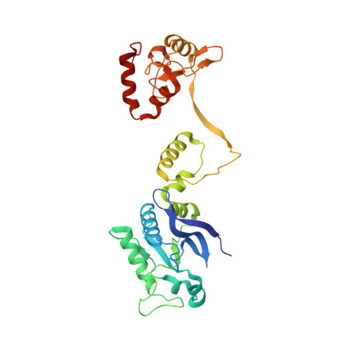
Activity and structure of EcoKMcrA.

Czapinska, H.Kowalska, M.Zagorskaite, E.Manakova, E.Slyvka, A.Xu, S.Y.Siksnys, V.Sasnauskas, G.Bochtler, M.

(2018) Nucleic Acids Res 46: 9829-9841

  • DOI: https://doi.org/10.1093/nar/gky731
  • Primary Citation Related Structures: 
    6GHC

  • PubMed Abstract: 

    Escherichia coli McrA (EcoKMcrA) acts as a methylcytosine and hydroxymethylcytosine dependent restriction endonuclease. We present a biochemical characterization of EcoKMcrA that includes the first demonstration of its endonuclease activity, small angle X-ray scattering (SAXS) data, and a crystal structure of the enzyme in the absence of DNA. Our data indicate that EcoKMcrA dimerizes via the anticipated C-terminal HNH domains, which together form a single DNA binding site. The N-terminal domains are not homologous to SRA domains, do not interact with each other, and have separate DNA binding sites. Electrophoretic mobility shift assay (EMSA) and footprinting experiments suggest that the N-terminal domains can sense the presence and sequence context of modified cytosines. Pyrrolocytosine fluorescence data indicate no base flipping. In vitro, EcoKMcrA DNA endonuclease activity requires Mn2+ ions, is not strictly methyl dependent, and is not observed when active site variants of the enzyme are used. In cells, EcoKMcrA specifically restricts DNA that is modified in the correct sequence context. This activity is impaired by mutations of the nuclease active site, unless the enzyme is highly overexpressed.


  • Organizational Affiliation
    • International Institute of Molecular and Cell Biology, Trojdena 4, 02-109 Warsaw, Poland.

Macromolecule Content 

  • Total Structure Weight: 65.71 kDa 
  • Atom Count: 4,577 
  • Modeled Residue Count: 569 
  • Deposited Residue Count: 574 
  • Unique protein chains: 1

Macromolecules

Find similar proteins by:|  3D Structure
Entity ID: 1
MoleculeChains  Sequence LengthOrganismDetailsImage
5-methylcytosine-specific restriction enzyme A
A, B
287Escherichia coli K-12Mutation(s): 0 
Gene Names: mcrArglAb1159JW1145
EC: 3.1.21
UniProt
Find proteins for P24200 (Escherichia coli (strain K12))
Explore P24200 
Go to UniProtKB:  P24200
Entity Groups
Sequence Clusters30% Identity50% Identity70% Identity90% Identity95% Identity100% Identity
UniProt GroupP24200
Sequence Annotations
Expand
Reference Sequence

Experimental Data & Validation

Experimental Data

  • Method: X-RAY DIFFRACTION
  • Resolution: 2.85 Å
  • R-Value Free:  0.283 (Depositor), 0.283 (DCC) 
  • R-Value Work:  0.218 (Depositor), 0.218 (DCC) 
  • R-Value Observed: 0.221 (Depositor) 
Space Group: P 21 21 21
Unit Cell:
Length ( Å )Angle ( ˚ )
a = 82.045α = 90
b = 90.383β = 90
c = 95.406γ = 90
Software Package:
Software NamePurpose
REFMACrefinement
XDSdata reduction
XDSdata scaling
SHELXDEphasing
autoSHARPphasing
DMphasing
BUCCANEERmodel building
PARROTphasing

Structure Validation

View Full Validation Report


Entry History 

Deposition Data

Revision History  (Full details and data files)

  • Version 1.0: 2018-08-08
    Type: Initial release
  • Version 1.1: 2018-08-22
    Changes: Data collection, Database references
  • Version 1.2: 2018-10-24
    Changes: Data collection, Database references
  • Version 1.3: 2024-05-15
    Changes: Data collection, Database references, Derived calculations