The Mechanism of Acetyl Transfer Catalyzed by Mycobacterium tuberculosis GlmU.
Craggs, P.D., Mouilleron, S., Rejzek, M., de Chiara, C., Young, R.J., Field, R.A., Argyrou, A., de Carvalho, L.P.S.(2018) Biochemistry 57: 3387-3401
- PubMed: 29684272 Search on PubMedSearch on PubMed Central
- DOI: https://doi.org/10.1021/acs.biochem.8b00121
- Primary Citation Related Structures:
6GE9 - PubMed Abstract:
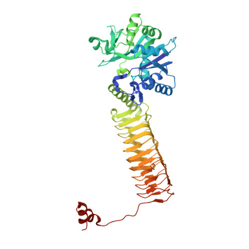
The biosynthetic pathway of peptidoglycan is essential for Mycobacterium tuberculosis. We report here the acetyltransferase substrate specificity and catalytic mechanism of the bifunctional N-acetyltransferase/uridylyltransferase from M. tuberculosis (GlmU). This enzyme is responsible for the final two steps of the synthesis of UDP- N-acetylglucosamine, which is an essential precursor of peptidoglycan, from glucosamine 1-phosphate, acetyl-coenzyme A, and uridine 5'-triphosphate. GlmU utilizes ternary complex formation to transfer an acetyl from acetyl-coenzyme A to glucosamine 1-phosphate to form N-acetylglucosamine 1-phosphate. Steady-state kinetic studies and equilibrium binding experiments indicate that GlmU follows a steady-state ordered kinetic mechanism, with acetyl-coenzyme A binding first, which triggers a conformational change in GlmU, followed by glucosamine 1-phosphate binding. Coenzyme A is the last product to dissociate. Chemistry is partially rate-limiting as indicated by pH-rate studies and solvent kinetic isotope effects. A novel crystal structure of a mimic of the Michaelis complex, with glucose 1-phosphate and acetyl-coenzyme A, helps us to propose the residues involved in deprotonation of glucosamine 1-phosphate and the loop movement that likely generates the active site required for glucosamine 1-phosphate to bind. Together, these results pave the way for the rational discovery of improved inhibitors against M. tuberculosis GlmU, some of which might become candidates for antibiotic discovery programs.
- Platform Technology and Science , GlaxoSmithKline , Stevenage , U.K.
Organizational Affiliation: