6G8E | pdb_00006g8e

Crystal Structure of the Amyloid-like VTQVGF segment from the R5 repeat of the E. coli Biofilm-associated CsgA Curli protein


Experimental Data Snapshot

  • Method: X-RAY DIFFRACTION
  • Resolution: 1.70 Å
  • R-Value Free: 
    0.132 (Depositor), 0.148 (DCC) 
  • R-Value Work: 
    0.123 (Depositor), 0.136 (DCC) 
  • R-Value Observed: 
    0.123 (Depositor) 

Starting Model: in silico
View more details

wwPDB Validation 3D Report Full Report

Validation slider image for 6G8E

This is version 1.3 of the entry. See complete history

Literature

Structural Insights into Curli CsgA Cross-beta Fibril Architecture Inspire Repurposing of Anti-amyloid Compounds as Anti-biofilm Agents.

Perov, S.Lidor, O.Salinas, N.Golan, N.Tayeb-Fligelman, E.Deshmukh, M.Willbold, D.Landau, M.

(2019) PLoS Pathog 15: e1007978-e1007978

  • DOI: https://doi.org/10.1371/journal.ppat.1007978
  • Primary Citation Related Structures: 
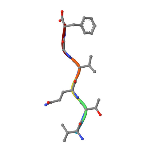
    6G8C, 6G8D, 6G8E, 6G9G

  • PubMed Abstract: 

    Curli amyloid fibrils secreted by Enterobacteriaceae mediate host cell adhesion and contribute to biofilm formation, thereby promoting bacterial resistance to environmental stressors. Here, we present crystal structures of amyloid-forming segments from the major curli subunit, CsgA, revealing steric zipper fibrils of tightly mated β-sheets, demonstrating a structural link between curli and human pathological amyloids. D-enantiomeric peptides, originally developed to interfere with Alzheimer's disease-associated amyloid-β, inhibited CsgA fibrillation and reduced biofilm formation in Salmonella typhimurium. Moreover, as previously shown, CsgA fibrils cross-seeded fibrillation of amyloid-β, providing support for the proposed structural resemblance and potential for cross-species amyloid interactions. The presented findings provide structural insights into amyloidogenic regions important for curli formation, suggest a novel strategy for disrupting amyloid-structured biofilms, and hypothesize on the formation of self-propagating prion-like species originating from a microbial source that could influence neurodegenerative diseases.


  • Organizational Affiliation
    • Department of Biology, Technion-Israel Institute of Technology, Haifa, Israel.

Macromolecule Content 

  • Total Structure Weight: 1.3 kDa 
  • Atom Count: 98 
  • Modeled Residue Count: 12 
  • Deposited Residue Count: 12 
  • Unique protein chains: 1

Macromolecules

Find similar proteins by:  Sequence   |   3D Structure  
Entity ID: 1
MoleculeChains  Sequence LengthOrganismDetailsImage
Major curlin subunitA [auth B],
B [auth A]
6Escherichia coli K-12Mutation(s): 0 
UniProt
Find proteins for P28307 (Escherichia coli (strain K12))
Explore P28307 
Go to UniProtKB:  P28307
Entity Groups
UniProt GroupP28307
Sequence Annotations
Expand
Reference Sequence

Experimental Data & Validation

Experimental Data

  • Method: X-RAY DIFFRACTION
  • Resolution: 1.70 Å
  • R-Value Free:  0.132 (Depositor), 0.148 (DCC) 
  • R-Value Work:  0.123 (Depositor), 0.136 (DCC) 
  • R-Value Observed: 0.123 (Depositor) 
Space Group: P 1
Unit Cell:
Length ( Å )Angle ( ˚ )
a = 4.81α = 65.82
b = 19.34β = 83.72
c = 21.9γ = 83.2
Software Package:
Software NamePurpose
XDSdata reduction
XSCALEdata scaling
PHASERphasing
REFMACrefinement
PDB_EXTRACTdata extraction

Structure Validation

View Full Validation Report


Entry History 

Deposition Data

Revision History  (Full details and data files)

  • Version 1.0: 2019-04-24
    Type: Initial release
  • Version 1.1: 2019-10-23
    Changes: Data collection, Database references
  • Version 1.2: 2019-12-25
    Changes: Data collection
  • Version 1.3: 2024-05-01
    Changes: Data collection, Database references, Refinement description