


Identifying small molecule binding sites for epigenetic proteins at domain-domain interfaces
Talon, R.P.H., Bowkett, D., Tallant, C., Schofield, C., von Delft, F., Knapp, S., Bruton, G., Brennan, P.E.(2018) bioRxiv
Experimental Data Snapshot
Starting Model: other
View more details
(2018) bioRxiv
Entity ID: 1 | |||||
|---|---|---|---|---|---|
| Molecule | Chains | Sequence Length | Organism | Details | Image |
| Nuclear autoantigen Sp-100 | 180 | Homo sapiens | Mutation(s): 0 Gene Names: SP100 | 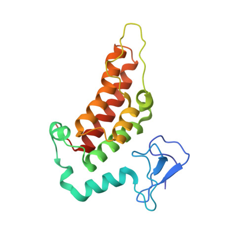 | |
UniProt & NIH Common Fund Data Resources | |||||
PHAROS: P23497 GTEx: ENSG00000067066 | |||||
Entity Groups | |||||
| Sequence Clusters | 30% Identity50% Identity70% Identity90% Identity95% Identity100% Identity | ||||
| UniProt Group | P23497 | ||||
Sequence AnnotationsExpand | |||||
Reference Sequence | |||||
| Ligands 5 Unique | |||||
|---|---|---|---|---|---|
| ID | Chains | Name / Formula / InChI Key | 2D Diagram | 3D Interactions | |
| MES Download:Ideal Coordinates CCD File | E [auth A] | 2-(N-MORPHOLINO)-ETHANESULFONIC ACID C6 H13 N O4 S SXGZJKUKBWWHRA-UHFFFAOYSA-N |  | ||
| ENK (Subject of Investigation/LOI) Download:Ideal Coordinates CCD File | H [auth A] | (3-phenyl-1,2,4-oxadiazol-5-yl)methanamine C9 H9 N3 O QFBMJBDECSEYCZ-UHFFFAOYSA-N |  | ||
| ZN Download:Ideal Coordinates CCD File | C [auth A], D [auth A], I [auth B], J [auth B] | ZINC ION Zn PTFCDOFLOPIGGS-UHFFFAOYSA-N |  | ||
| EDO Download:Ideal Coordinates CCD File | F [auth A], G [auth A], K [auth B], L [auth B] | 1,2-ETHANEDIOL C2 H6 O2 LYCAIKOWRPUZTN-UHFFFAOYSA-N |  | ||
| CL Download:Ideal Coordinates CCD File | M [auth B] | CHLORIDE ION Cl VEXZGXHMUGYJMC-UHFFFAOYSA-M |  | ||
| Length ( Å ) | Angle ( ˚ ) |
|---|---|
| a = 127.879 | α = 90 |
| b = 45.426 | β = 102.14 |
| c = 83.477 | γ = 90 |
| Software Name | Purpose |
|---|---|
| PHENIX | refinement |
| XDS | data reduction |
| Aimless | data scaling |
| PHASER | phasing |
| Funding Organization | Location | Grant Number |
|---|---|---|
| United Kingdom | -- |


