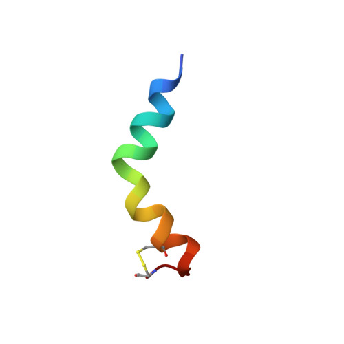
Structural and positional studies of the antimicrobial peptide brevinin-1BYa in membrane-mimetic environments.
Timmons, P.B., O'Flynn, D., Conlon, J.M., Hewage, C.M.(2019) J Pept Sci 25: e3208-e3208
- PubMed: 31721374
- DOI: https://doi.org/10.1002/psc.3208
- Primary Citation of Related Structures:
6G4I, 6G4K, 6G4U - PubMed Abstract:
Brevinin-1BYa (FLPILASLAAKFGPKLFCLVTKKC), first isolated from skin secretions of the foothill yellow-legged frog Rana boylii, shows broad-spectrum activity, being particularly effective against opportunistic yeast pathogens. The structure of brevinin-1BYa was investigated in various solution and membrane-mimicking environments by proton nuclear magnetic resonance ( 1 H-NMR) spectroscopy and molecular modelling. The peptide does not possess a secondary structure in aqueous solution. In a 33% 2,2,2-trifluoroethanol (TFE-d 3 )-H 2 O solvent mixture, as well as in membrane-mimicking sodium dodecyl sulfate and dodecylphosphocholine micelles, the peptide's structure is characterised by a flexible helix-hinge-helix motif, with the hinge located at the Gly 13 /Pro 14 residues, and the two α-helices extending from Pro 3 to Phe 12 and from Pro 14 to Thr 21 . Positional studies involving the peptide in sodium dodecyl sulfate and dodecylphosphocholine micelles using 5-doxyl-labelled stearic acid and manganese chloride paramagnetic probes show that the peptide's helical segments lie parallel to the micellar surface, with the residues on the hydrophobic face of the amphipathic helices facing towards the micelle core and the hydrophilic residues pointing outwards, suggesting that the peptide exerts its biological activity by a non-pore-forming mechanism.
- UCD School of Biomolecular and Biomedical Science, UCD Centre for Synthesis and Chemical Biology, UCD Conway Institute, University College Dublin, Dublin, Ireland.
Organizational Affiliation:


