6G46 | pdb_00006g46

Red kidney bean purple acid phosphatase in complex with 2-(Naphthalen-1-yl)thiazole-4-carboxylic acid


Experimental Data Snapshot

  • Method: X-RAY DIFFRACTION
  • Resolution: 2.40 Å
  • R-Value Free: 
    0.223 (Depositor), 0.223 (DCC) 
  • R-Value Work: 
    0.172 (Depositor), 0.173 (DCC) 
  • R-Value Observed: 
    0.175 (Depositor) 

Starting Model: experimental
View more details

wwPDB Validation 3D Report Full Report

Validation slider image for 6G46

Ligand Structure Quality Assessment 


This is version 2.2 of the entry. See complete history

Literature

Purple acid phosphatase inhibitors as leads for osteoporosis chemotherapeutics.

Hussein, W.M.Feder, D.Schenk, G.Guddat, L.W.McGeary, R.P.

(2018) Eur J Med Chem 157: 462-479

  • DOI: https://doi.org/10.1016/j.ejmech.2018.08.004
  • Primary Citation Related Structures: 
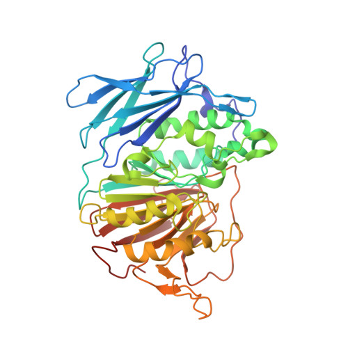
    6G46

  • PubMed Abstract: 

    Purple acid phosphatases (PAPs) are metalloenzymes that catalyse the hydrolysis of phosphate esters under acidic conditions. Their active site contains a Fe(III)Fe(II) metal centre in mammals and a Fe(III)Zn(II) or Fe(III)Mn(II) metal centre in plants. In humans, elevated PAP levels in serum strongly correlate with the progression of osteoporosis and metabolic bone malignancies, which make PAP a target suitable for the development of chemotherapeutics to combat bone ailments. Due to difficulties in obtaining the human enzyme, the corresponding enzymes from red kidney bean and pig have been used previously to develop specific PAP inhibitors. Here, existing lead compounds were further elaborated to create a series of inhibitors with K i values as low as ∼30 μM. The inhibition constants of these compounds were of comparable magnitude for pig and red kidney bean PAPs, indicating that relevant binding interactions are conserved. The crystal structure of red kidney bean PAP in complex with the most potent inhibitor in this series, compound 4f, was solved to 2.40 Å resolution. This inhibitor coordinates directly to the binuclear metal centre in the active site as expected based on its competitive mode of inhibition. Docking simulations predict that this compound binds to human PAP in a similar mode. This study presents the first example of a PAP structure in complex with an inhibitor that is of relevance to the development of anti-osteoporotic chemotherapeutics.


  • Organizational Affiliation
    • The University of Queensland, School of Chemistry and Molecular Biosciences, Brisbane, QLD 4072, Australia; Helwan University, Pharmaceutical Organic Chemistry Department, Faculty of Pharmacy, Ein Helwan, Helwan, Egypt.

Macromolecule Content 

  • Total Structure Weight: 206.72 kDa 
  • Atom Count: 15,764 
  • Modeled Residue Count: 1,696 
  • Deposited Residue Count: 1,704 
  • Unique protein chains: 1

Macromolecules

Find similar proteins by:|  3D Structure
Entity ID: 1
MoleculeChains  Sequence LengthOrganismDetailsImage
Fe(3+)-Zn(2+) purple acid phosphataseA,
B,
C [auth D],
D [auth C]
426Phaseolus vulgarisMutation(s): 0 
EC: 3.1.3.2
UniProt
Find proteins for P80366 (Phaseolus vulgaris)
Explore P80366 
Go to UniProtKB:  P80366
Entity Groups
Sequence Clusters30% Identity50% Identity70% Identity90% Identity95% Identity100% Identity
UniProt GroupP80366
Glycosylation
Glycosylation Sites: 4
Sequence Annotations
Expand
Reference Sequence

Oligosaccharides

Help  
Entity ID: 2
MoleculeChains Length2D Diagram GlycosylationD Interactions
alpha-L-fucopyranose-(1-3)-[2-acetamido-2-deoxy-beta-D-glucopyranose-(1-4)]2-acetamido-2-deoxy-beta-D-glucopyranose
E, F, G, I, J
E, F, G, I, J, M
3N-Glycosylation
Glycosylation Resources
GlyTouCan: G11971MR
GlyCosmos: G11971MR
GlyGen: G11971MR
Entity ID: 3
MoleculeChains Length2D Diagram GlycosylationD Interactions
alpha-L-fucopyranose-(1-3)-2-acetamido-2-deoxy-beta-D-glucopyranose
H, K, L
2N-Glycosylation
Glycosylation Resources
GlyTouCan: G80587NA
GlyCosmos: G80587NA
GlyGen: G80587NA

Small Molecules

Ligands 9 Unique
IDChains Name / Formula / InChI Key2D Diagram3D Interactions
ELH

Query on ELH


Download:Ideal Coordinates CCD File
AA [auth B],
S [auth A]
2-naphthalen-1-yl-1,3-thiazole-4-carboxylic acid
C14 H9 N O2 S
AQRUWPVKXHZCKR-UHFFFAOYSA-N
NAG

Query on NAG


Download:Ideal Coordinates CCD File
KA [auth D]
MA [auth D]
R [auth A]
SA [auth C]
TA [auth C]
KA [auth D],
MA [auth D],
R [auth A],
SA [auth C],
TA [auth C],
X [auth B],
Y [auth B]
2-acetamido-2-deoxy-beta-D-glucopyranose
C8 H15 N O6
OVRNDRQMDRJTHS-FMDGEEDCSA-N
SO4

Query on SO4


Download:Ideal Coordinates CCD File
JA [auth D]
NA [auth D]
P [auth A]
Q [auth A]
UA [auth C]
JA [auth D],
NA [auth D],
P [auth A],
Q [auth A],
UA [auth C],
VA [auth C],
W [auth B],
Z [auth B]
SULFATE ION
O4 S
QAOWNCQODCNURD-UHFFFAOYSA-L
GOL

Query on GOL


Download:Ideal Coordinates CCD File
XA [auth C]GLYCEROL
C3 H8 O3
PEDCQBHIVMGVHV-UHFFFAOYSA-N
ZN

Query on ZN


Download:Ideal Coordinates CCD File
HA [auth D],
N [auth A],
QA [auth C],
U [auth B]
ZINC ION
Zn
PTFCDOFLOPIGGS-UHFFFAOYSA-N
EDO

Query on EDO


Download:Ideal Coordinates CCD File
EA [auth B]
FA [auth B]
GA [auth B]
LA [auth D]
PA [auth D]
EA [auth B],
FA [auth B],
GA [auth B],
LA [auth D],
PA [auth D],
T [auth A],
WA [auth C]
1,2-ETHANEDIOL
C2 H6 O2
LYCAIKOWRPUZTN-UHFFFAOYSA-N
IPA

Query on IPA


Download:Ideal Coordinates CCD File
DA [auth B]ISOPROPYL ALCOHOL
C3 H8 O
KFZMGEQAYNKOFK-UHFFFAOYSA-N
FE

Query on FE


Download:Ideal Coordinates CCD File
IA [auth D],
O [auth A],
RA [auth C],
V [auth B]
FE (III) ION
Fe
VTLYFUHAOXGGBS-UHFFFAOYSA-N
NA

Query on NA


Download:Ideal Coordinates CCD File
BA [auth B],
CA [auth B],
OA [auth D]
SODIUM ION
Na
FKNQFGJONOIPTF-UHFFFAOYSA-N

Experimental Data & Validation

Experimental Data

  • Method: X-RAY DIFFRACTION
  • Resolution: 2.40 Å
  • R-Value Free:  0.223 (Depositor), 0.223 (DCC) 
  • R-Value Work:  0.172 (Depositor), 0.173 (DCC) 
  • R-Value Observed: 0.175 (Depositor) 
Space Group: P 31 2 1
Unit Cell:
Length ( Å )Angle ( ˚ )
a = 126.208α = 90
b = 126.208β = 90
c = 297.119γ = 120
Software Package:
Software NamePurpose
PHENIXrefinement
CrystalCleardata reduction
pointlessdata scaling
PHASERphasing

Structure Validation

View Full Validation Report


Ligand Structure Quality Assessment 


Entry History 

& Funding Information

Deposition Data

  • Released Date: 2018-09-26 
  • Deposition Author(s): Feder, D.F.

Funding OrganizationLocationGrant Number
Australian Research CouncilAustraliaDP0986292
Australian Research CouncilAustraliaDP150104358

Revision History  (Full details and data files)

  • Version 1.0: 2018-09-26
    Type: Initial release
  • Version 2.0: 2020-07-29
    Type: Remediation
    Reason: Carbohydrate remediation
    Changes: Advisory, Atomic model, Data collection, Derived calculations, Structure summary
  • Version 2.1: 2024-01-17
    Changes: Advisory, Data collection, Database references, Derived calculations, Refinement description, Structure summary
  • Version 2.2: 2024-11-20
    Changes: Structure summary