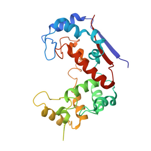
6FXO

Crystal structure of Major Bifunctional Autolysin


Experimental Data Snapshot

  • Method: X-RAY DIFFRACTION
  • Resolution: 2.50 Å
  • R-Value Free: 0.330 
  • R-Value Work: 0.295 
  • R-Value Observed: 0.295 

Starting Model: other
View more details

wwPDB Validation   3D Report Full Report


This is version 2.2 of the entry. See complete history


Literature

Domain sliding of two Staphylococcus aureus N-acetylglucosaminidases enables their substrate-binding prior to its catalysis.

Pintar, S.Borisek, J.Usenik, A.Perdih, A.Turk, D.

(2020) Commun Biol 3: 178-178

  • DOI: https://doi.org/10.1038/s42003-020-0911-7
  • Primary Citation of Related Structures:  
    6FXO, 6FXP

  • PubMed Abstract: 

    To achieve productive binding, enzymes and substrates must align their geometries to complement each other along an entire substrate binding site, which may require enzyme flexibility. In pursuit of novel drug targets for the human pathogen S. aureus, we studied peptidoglycan N-acetylglucosaminidases, whose structures are composed of two domains forming a V-shaped active site cleft. Combined insights from crystal structures supported by site-directed mutagenesis, modeling, and molecular dynamics enabled us to elucidate the substrate binding mechanism of SagB and AtlA-gl. This mechanism requires domain sliding from the open form observed in their crystal structures, leading to polysaccharide substrate binding in the closed form, which can enzymatically process the bound substrate. We suggest that these two hydrolases must exhibit unusual extents of flexibility to cleave the rigid structure of a bacterial cell wall.


  • Organizational Affiliation

    Department of Biochemistry, Molecular and Structural Biology, Jožef Stefan Institute, Jamova cesta 39, 1000, Ljubljana, Slovenia.


Macromolecules
Find similar proteins by:  (by identity cutoff)  |  3D Structure
Entity ID: 1
MoleculeChains Sequence LengthOrganismDetailsImage
Bifunctional autolysin244Staphylococcus aureus subsp. aureus Mu50Mutation(s): 0 
Gene Names: atlnagSAV1052
EC: 3.5.1.28 (PDB Primary Data), 3.2.1.96 (PDB Primary Data)
UniProt
Find proteins for Q931U5 (Staphylococcus aureus (strain Mu50 / ATCC 700699))
Explore Q931U5 
Go to UniProtKB:  Q931U5
Entity Groups  
Sequence Clusters30% Identity50% Identity70% Identity90% Identity95% Identity100% Identity
UniProt GroupQ931U5
Sequence Annotations
Expand
  • Reference Sequence
Experimental Data & Validation

Experimental Data

  • Method: X-RAY DIFFRACTION
  • Resolution: 2.50 Å
  • R-Value Free: 0.330 
  • R-Value Work: 0.295 
  • R-Value Observed: 0.295 
  • Space Group: P 64 2 2
Unit Cell:
Length ( Å )Angle ( ˚ )
a = 110.168α = 90
b = 110.168β = 90
c = 92.39γ = 120
Software Package:
Software NamePurpose
MAINrefinement
XDSdata reduction
XDSdata scaling
PHASERphasing

Structure Validation

View Full Validation Report


Entry History & Funding Information

Deposition Data


Funding OrganizationLocationGrant Number
Slovenian Research AgencySloveniaP1-0048

Revision History  (Full details and data files)

  • Version 1.0: 2019-03-20
    Type: Initial release
  • Version 2.0: 2020-07-22
    Type: Coordinate replacement
    Reason: Polymer geometry
    Changes: Advisory, Atomic model, Author supporting evidence, Data collection, Database references, Derived calculations, Other, Polymer sequence, Refinement description, Source and taxonomy, Structure summary
  • Version 2.1: 2020-09-30
    Changes: Database references
  • Version 2.2: 2024-05-01
    Changes: Advisory, Data collection, Database references, Refinement description