6FXM

Crystal Structure of full-length Human Lysyl Hydroxylase LH3 - Cocrystal with Mn2+


Experimental Data Snapshot

  • Method: X-RAY DIFFRACTION
  • Resolution: 2.10 Å
  • R-Value Free: 0.252 
  • R-Value Work: 0.229 
  • R-Value Observed: 0.230 

wwPDB Validation   3D Report Full Report


Ligand Structure Quality Assessment 


This is version 2.0 of the entry. See complete history


Literature

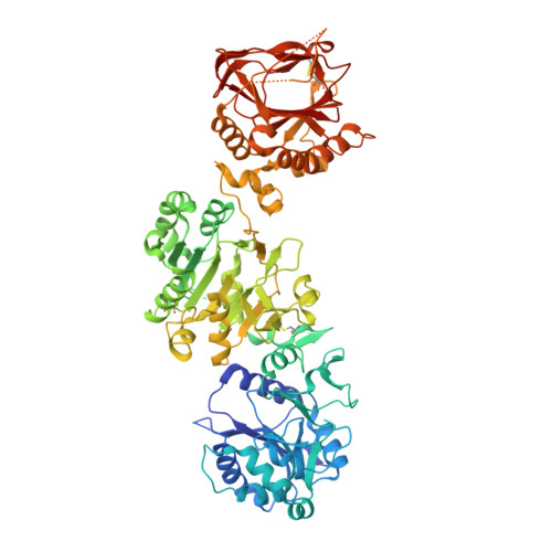
Molecular architecture of the multifunctional collagen lysyl hydroxylase and glycosyltransferase LH3.

Scietti, L.Chiapparino, A.De Giorgi, F.Fumagalli, M.Khoriauli, L.Nergadze, S.Basu, S.Olieric, V.Cucca, L.Banushi, B.Profumo, A.Giulotto, E.Gissen, P.Forneris, F.

(2018) Nat Commun 9: 3163-3163

  • DOI: https://doi.org/10.1038/s41467-018-05631-5
  • Primary Citation of Related Structures:  
    6FXK, 6FXM, 6FXR, 6FXT, 6FXX, 6FXY

  • PubMed Abstract: 

    Lysyl hydroxylases catalyze hydroxylation of collagen lysines, and sustain essential roles in extracellular matrix (ECM) maturation and remodeling. Malfunctions in these enzymes cause severe connective tissue disorders. Human lysyl hydroxylase 3 (LH3/PLOD3) bears multiple enzymatic activities, as it catalyzes collagen lysine hydroxylation and also their subsequent glycosylation. Our understanding of LH3 functions is currently hampered by lack of molecular structure information. Here, we present high resolution crystal structures of full-length human LH3 in complex with cofactors and donor substrates. The elongated homodimeric LH3 architecture shows two distinct catalytic sites at the N- and C-terminal boundaries of each monomer, separated by an accessory domain. The glycosyltransferase domain displays distinguishing features compared to other known glycosyltransferases. Known disease-related mutations map in close proximity to the catalytic sites. Collectively, our results provide a structural framework characterizing the multiple functions of LH3, and the molecular mechanisms of collagen-related diseases involving human lysyl hydroxylases.


  • Organizational Affiliation

    The Armenise-Harvard Laboratory of Structural Biology, Department of Biology and Biotechnology, University of Pavia, Via Ferrata 9/A, 27100, Pavia, Italy.


Macromolecules
Find similar proteins by:  (by identity cutoff)  |  3D Structure
Entity ID: 1
MoleculeChains Sequence LengthOrganismDetailsImage
Procollagen-lysine,2-oxoglutarate 5-dioxygenase 3718Homo sapiensMutation(s): 0 
Gene Names: PLOD3
EC: 1.14.11.4
UniProt & NIH Common Fund Data Resources
Find proteins for O60568 (Homo sapiens)
Explore O60568 
Go to UniProtKB:  O60568
PHAROS:  O60568
GTEx:  ENSG00000106397 
Entity Groups  
Sequence Clusters30% Identity50% Identity70% Identity90% Identity95% Identity100% Identity
UniProt GroupO60568
Sequence Annotations
Expand
  • Reference Sequence
Oligosaccharides

Help

Entity ID: 2
MoleculeChains Length2D Diagram Glycosylation3D Interactions
2-acetamido-2-deoxy-beta-D-glucopyranose-(1-4)-2-acetamido-2-deoxy-beta-D-glucopyranose
B
2N-Glycosylation
Glycosylation Resources
GlyTouCan:  G42666HT
GlyCosmos:  G42666HT
GlyGen:  G42666HT
Small Molecules
Ligands 5 Unique
IDChains Name / Formula / InChI Key2D Diagram3D Interactions
NAG
Query on NAG

Download Ideal Coordinates CCD File 
K [auth A]2-acetamido-2-deoxy-beta-D-glucopyranose
C8 H15 N O6
OVRNDRQMDRJTHS-FMDGEEDCSA-N
AKG (Subject of Investigation/LOI)
Query on AKG

Download Ideal Coordinates CCD File 
G [auth A]2-OXOGLUTARIC ACID
C5 H6 O5
KPGXRSRHYNQIFN-UHFFFAOYSA-N
GOL
Query on GOL

Download Ideal Coordinates CCD File 
C [auth A],
D [auth A],
E [auth A],
F [auth A]
GLYCEROL
C3 H8 O3
PEDCQBHIVMGVHV-UHFFFAOYSA-N
FE2 (Subject of Investigation/LOI)
Query on FE2

Download Ideal Coordinates CCD File 
H [auth A]FE (II) ION
Fe
CWYNVVGOOAEACU-UHFFFAOYSA-N
MN (Subject of Investigation/LOI)
Query on MN

Download Ideal Coordinates CCD File 
I [auth A],
J [auth A]
MANGANESE (II) ION
Mn
WAEMQWOKJMHJLA-UHFFFAOYSA-N
Experimental Data & Validation

Experimental Data

  • Method: X-RAY DIFFRACTION
  • Resolution: 2.10 Å
  • R-Value Free: 0.252 
  • R-Value Work: 0.229 
  • R-Value Observed: 0.230 
  • Space Group: C 2 2 21
Unit Cell:
Length ( Å )Angle ( ˚ )
a = 97.27α = 90
b = 99.949β = 90
c = 225.138γ = 90
Software Package:
Software NamePurpose
PHENIXrefinement
XDSdata reduction
Aimlessdata scaling
SHARPphasing

Structure Validation

View Full Validation Report


Ligand Structure Quality Assessment 


Entry History & Funding Information

Deposition Data


Funding OrganizationLocationGrant Number
The Giovanni Armenise-Harvard FoundationUnited StatesCareer Development Award
Cariplo FoundationItaly2015-0768 - COME TRUE
AIRCItaly20075
European Commission MSCA-IFItaly745934 - COTETHERS
European Union - BIOSTRUCTItaly7551, 10205

Revision History  (Full details and data files)

  • Version 1.0: 2018-08-22
    Type: Initial release
  • Version 2.0: 2020-07-29
    Type: Remediation
    Reason: Carbohydrate remediation
    Changes: Atomic model, Data collection, Derived calculations, Structure summary