


Crystal structure of T. brucei PDE-B1 catalytic domain with inhibitor NPD-048
de Heuvel, E., Singh, A.K., Brown, D.G.To be published.
Experimental Data Snapshot
Starting Model: experimental
View more details
Entity ID: 1 | |||||
|---|---|---|---|---|---|
| Molecule | Chains | Sequence Length | Organism | Details | Image |
| Phosphodiesterase | 360 | Trypanosoma brucei | Mutation(s): 0 Gene Names: PDEB1 EC: 3.1.4 | 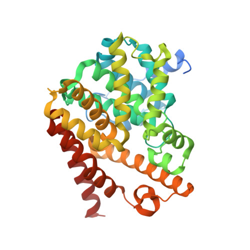 | |
UniProt | |||||
Entity Groups | |||||
| Sequence Clusters | 30% Identity50% Identity70% Identity90% Identity95% Identity100% Identity | ||||
| UniProt Group | Q8WQX9 | ||||
Sequence AnnotationsExpand | |||||
Reference Sequence | |||||
| Ligands 6 Unique | |||||
|---|---|---|---|---|---|
| ID | Chains | Name / Formula / InChI Key | 2D Diagram | 3D Interactions | |
| E6Z (Subject of Investigation/LOI) Download:Ideal Coordinates CCD File | BA [auth A], PA [auth B] | 3-{5-[(4aR,8aS)-3-cycloheptyl-4-oxo-3,4,4a,5,8,8a-hexahydrophthalazin-1-yl]-2-methoxyphenyl}prop-2-ynamide C25 H29 N3 O3 YALZXZYFCQHHIY-LEWJYISDSA-N |  | ||
| ZN Download:Ideal Coordinates CCD File | D [auth A], EA [auth B] | ZINC ION Zn PTFCDOFLOPIGGS-UHFFFAOYSA-N |  | ||
| EDO Download:Ideal Coordinates CCD File | E [auth A] F [auth A] FA [auth B] G [auth A] GA [auth B] | 1,2-ETHANEDIOL C2 H6 O2 LYCAIKOWRPUZTN-UHFFFAOYSA-N |  | ||
| GAI Download:Ideal Coordinates CCD File | MA [auth B] NA [auth B] Q [auth A] R [auth A] S [auth A] | GUANIDINE C H5 N3 ZRALSGWEFCBTJO-UHFFFAOYSA-N |  | ||
| FMT Download:Ideal Coordinates CCD File | AA [auth A], CA [auth A], OA [auth B], Y [auth A], Z [auth A] | FORMIC ACID C H2 O2 BDAGIHXWWSANSR-UHFFFAOYSA-N |  | ||
| MG Download:Ideal Coordinates CCD File | C [auth A], DA [auth B] | MAGNESIUM ION Mg JLVVSXFLKOJNIY-UHFFFAOYSA-N |  | ||
| Length ( Å ) | Angle ( ˚ ) |
|---|---|
| a = 149.052 | α = 90 |
| b = 114.73 | β = 109.43 |
| c = 63.8 | γ = 90 |
| Software Name | Purpose |
|---|---|
| REFMAC | refinement |
| MOSFLM | data reduction |
| Aimless | data scaling |
| PHASER | phasing |


