6FPJ

Structure of the AMPAR GluA3 N-terminal domain bound to phosphate


Experimental Data Snapshot

  • Method: X-RAY DIFFRACTION
  • Resolution: 1.96 Å
  • R-Value Free: 0.219 
  • R-Value Work: 0.179 
  • R-Value Observed: 0.181 

wwPDB Validation   3D Report Full Report


Ligand Structure Quality Assessment 


This is version 1.3 of the entry. See complete history


Literature

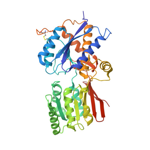
Druggability Simulations and X-Ray Crystallography Reveal a Ligand-Binding Site in the GluA3 AMPA Receptor N-Terminal Domain.

Lee, J.Y.Krieger, J.Herguedas, B.Garcia-Nafria, J.Dutta, A.Shaikh, S.A.Greger, I.H.Bahar, I.

(2019) Structure 27: 241-252.e3

  • DOI: https://doi.org/10.1016/j.str.2018.10.017
  • Primary Citation of Related Structures:  
    6FLR, 6FPJ

  • PubMed Abstract: 

    Ionotropic glutamate receptors (iGluRs) mediate the majority of excitatory neurotransmission in the brain. Their dysfunction is implicated in many neurological disorders, rendering iGluRs potential drug targets. Here, we performed a systematic analysis of the druggability of two major iGluR subfamilies, using molecular dynamics simulations in the presence of drug-like molecules. We demonstrate the applicability of druggability simulations by faithfully identifying known agonist and modulator sites on AMPA receptors (AMPARs) and NMDA receptors. Simulations produced the expected allosteric changes of the AMPAR ligand-binding domain in response to agonist. We also identified a novel ligand-binding site specific to the GluA3 AMPAR N-terminal domain (NTD), resulting from its unique conformational flexibility that we explored further with crystal structures trapped in vastly different states. In addition to providing an in-depth analysis into iGluR NTD dynamics, our approach identifies druggable sites and permits the determination of pharmacophoric features toward novel iGluR modulators.


  • Organizational Affiliation

    Department of Computational and Systems Biology, School of Medicine, University of Pittsburgh, 3501 Fifth Avenue, Suite 3064 BST3, Pittsburgh, PA 15260, USA.


Macromolecules
Find similar proteins by:  (by identity cutoff)  |  3D Structure
Entity ID: 1
MoleculeChains Sequence LengthOrganismDetailsImage
Glutamate receptor 3
A, B, C
390Rattus norvegicusMutation(s): 0 
Gene Names: Gria3Glur3
UniProt
Find proteins for P19492 (Rattus norvegicus)
Explore P19492 
Go to UniProtKB:  P19492
Entity Groups  
Sequence Clusters30% Identity50% Identity70% Identity90% Identity95% Identity100% Identity
UniProt GroupP19492
Sequence Annotations
Expand
  • Reference Sequence
Small Molecules
Ligands 4 Unique
IDChains Name / Formula / InChI Key2D Diagram3D Interactions
NAG
Query on NAG

Download Ideal Coordinates CCD File 
D [auth A]
E [auth A]
F [auth A]
K [auth B]
L [auth B]
D [auth A],
E [auth A],
F [auth A],
K [auth B],
L [auth B],
M [auth B],
V [auth C],
W [auth C]
2-acetamido-2-deoxy-beta-D-glucopyranose
C8 H15 N O6
OVRNDRQMDRJTHS-FMDGEEDCSA-N
PO4
Query on PO4

Download Ideal Coordinates CCD File 
G [auth A]
N [auth B]
O [auth B]
P [auth B]
Q [auth B]
G [auth A],
N [auth B],
O [auth B],
P [auth B],
Q [auth B],
R [auth B],
X [auth C]
PHOSPHATE ION
O4 P
NBIIXXVUZAFLBC-UHFFFAOYSA-K
GOL
Query on GOL

Download Ideal Coordinates CCD File 
U [auth B]GLYCEROL
C3 H8 O3
PEDCQBHIVMGVHV-UHFFFAOYSA-N
DMS
Query on DMS

Download Ideal Coordinates CCD File 
H [auth A]
I [auth A]
J [auth A]
S [auth B]
T [auth B]
H [auth A],
I [auth A],
J [auth A],
S [auth B],
T [auth B],
Y [auth C],
Z [auth C]
DIMETHYL SULFOXIDE
C2 H6 O S
IAZDPXIOMUYVGZ-UHFFFAOYSA-N
Experimental Data & Validation

Experimental Data

  • Method: X-RAY DIFFRACTION
  • Resolution: 1.96 Å
  • R-Value Free: 0.219 
  • R-Value Work: 0.179 
  • R-Value Observed: 0.181 
  • Space Group: P 21 21 2
Unit Cell:
Length ( Å )Angle ( ˚ )
a = 109.15α = 90
b = 164.77β = 90
c = 63.35γ = 90
Software Package:
Software NamePurpose
REFMACrefinement
PDB_EXTRACTdata extraction
xia2data reduction
xia2data scaling
MOLREPphasing

Structure Validation

View Full Validation Report


Ligand Structure Quality Assessment 


Entry History 

Deposition Data

Revision History  (Full details and data files)

  • Version 1.0: 2018-12-19
    Type: Initial release
  • Version 1.1: 2019-02-13
    Changes: Data collection, Database references
  • Version 1.2: 2020-07-29
    Type: Remediation
    Reason: Carbohydrate remediation
    Changes: Data collection, Derived calculations, Structure summary
  • Version 1.3: 2024-01-17
    Changes: Data collection, Database references, Refinement description, Structure summary