


Structure of the Mannose Transporter IIA Domain from Streptococcus pneumoniae
Magoch, M., Grudnik, P., Nogly, P., Dubin, G., Archer, M., Ma, P.To be published.
Experimental Data Snapshot
Starting Model: experimental
View more details
wwPDB Validation 3D Report Full Report

Entity ID: 1 | |||||
|---|---|---|---|---|---|
| Molecule | Chains | Sequence Length | Organism | Details | Image |
| PTS system mannose-specific transporter subunit IIAB | 140 | Streptococcus pneumoniae | Mutation(s): 0 Gene Names: manL_2, ERS020456_01132 EC: 2.7.1.69 | 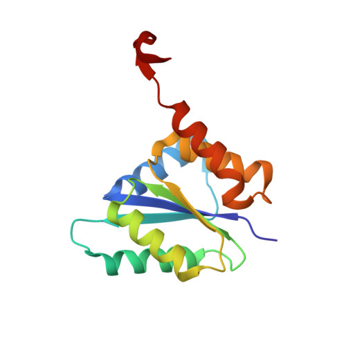 | |
| Length ( Å ) | Angle ( ˚ ) |
|---|---|
| a = 138.528 | α = 90 |
| b = 138.528 | β = 90 |
| c = 69.767 | γ = 120 |
| Software Name | Purpose |
|---|---|
| PHENIX | refinement |
| PDB_EXTRACT | data extraction |
| XDS | data reduction |
| SCALA | data scaling |
| PHASER | phasing |
| Coot | model building |


