A Fluorescent Probe Identifies Active Site Ligands of Inositol Pentakisphosphate 2-Kinase.
Whitfield, H., Gilmartin, M., Baker, K., Riley, A.M., Godage, H.Y., Potter, B.V.L., Hemmings, A.M., Brearley, C.A.(2018) J Med Chem 61: 8838-8846
- PubMed: 30160967 Search on PubMed
- DOI: https://doi.org/10.1021/acs.jmedchem.8b01022
- Primary Citation Related Structures:
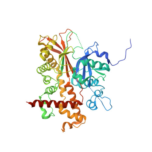
6FJK, 6FL3, 6FL8, 6GFG, 6GFH - PubMed Abstract:
Inositol pentakisphosphate 2-kinase catalyzes the phosphorylation of the axial 2-OH of myo-inositol 1,3,4,5,6-pentakisphosphate for de novo synthesis of myo-inositol hexakisphosphate. Disruption of inositol pentakisphosphate 2-kinase profoundly influences cellular processes, from nuclear mRNA export and phosphate homeostasis in yeast and plants to establishment of left-right asymmetry in zebrafish. We elaborate an active site fluorescent probe that allows high throughput screening of Arabidopsis inositol pentakisphosphate 2-kinase. We show that the probe has a binding constant comparable to the K m values of inositol phosphate substrates of this enzyme and can be used to prospect for novel substrates and inhibitors of inositol phosphate kinases. We identify several micromolar K i inhibitors and validate this approach by solving the crystal structure of protein in complex with purpurogallin. We additionally solve structures of protein in complexes with epimeric higher inositol phosphates. This probe may find utility in characterization of a wide family of inositol phosphate kinases.
- School of Biological Sciences , University of East Anglia , Norwich Research Park , Norwich NR4 7TJ , U.K.
Organizational Affiliation: