6FIH | pdb_00006fih

Crystal structure of the ANX2 ectodomain from Arabidopsis thaliana


Experimental Data Snapshot

  • Method: X-RAY DIFFRACTION
  • Resolution: 1.08 Å
  • R-Value Free: 
    0.183 (Depositor), 0.182 (DCC) 
  • R-Value Work: 
    0.159 (Depositor), 0.159 (DCC) 
  • R-Value Observed: 
    0.160 (Depositor) 

wwPDB Validation 3D Report Full Report

Validation slider image for 6FIH

Ligand Structure Quality Assessment 


This is version 2.1 of the entry. See complete history

Literature

Crystal structures of two tandem malectin-like receptor kinases involved in plant reproduction.

Moussu, S.Augustin, S.Roman, A.O.Broyart, C.Santiago, J.

(2018) Acta Crystallogr D Struct Biol 74: 671-680

  • DOI: https://doi.org/10.1107/S205979831800774X
  • Primary Citation Related Structures: 
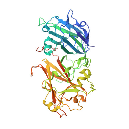
    6FIG, 6FIH

  • PubMed Abstract: 

    Complex cell-to-cell communication between the male pollen tube and the female reproductive organs is required for plant fertilization. A family of Catharanthus roseus receptor kinase 1-like (CrRLK1L) membrane receptors has been genetically implicated in this process. Here, crystal structures of the CrRLK1Ls ANXUR1 and ANXUR2 are reported at 1.48 and 1.1 Å resolution, respectively. The structures reveal a novel arrangement of two malectin-like domains connected by a short β-hairpin linker and stabilized by calcium ions. The canonical carbohydrate-interaction surfaces of related animal and bacterial carbohydrate-binding modules are not conserved in plant CrRLK1Ls. In line with this, the binding of chemically diverse oligosaccharides to ANXUR1 and HERCULES1 could not be detected. Instead, CrRLK1Ls have evolved a protein-protein interface between their malectin domains which forms a deep cleft lined by highly conserved aromatic and polar residues. Analysis of the glycosylation patterns of different CrRLK1Ls and their oligomeric states suggests that this cleft could resemble a binding site for a ligand required for receptor activation of CrRLK1Ls.


  • Organizational Affiliation
    • The Plant Signaling Mechanisms Laboratory, Department of Plant Molecular Biology, University of Lausanne, 1015 Lausanne, Switzerland.

Macromolecule Content 

  • Total Structure Weight: 47.56 kDa 
  • Atom Count: 3,574 
  • Modeled Residue Count: 379 
  • Deposited Residue Count: 412 
  • Unique protein chains: 1

Macromolecules

Find similar proteins by:|  3D Structure
Entity ID: 1
MoleculeChains  Sequence LengthOrganismDetailsImage
Receptor-like protein kinase ANXUR2412Arabidopsis thalianaMutation(s): 0 
Gene Names: ANX2At5g28680F4I4.60
EC: 2.7.11.1
UniProt
Find proteins for Q3E8W4 (Arabidopsis thaliana)
Explore Q3E8W4 
Go to UniProtKB:  Q3E8W4
Entity Groups
Sequence Clusters30% Identity50% Identity70% Identity90% Identity95% Identity100% Identity
UniProt GroupQ3E8W4
Glycosylation
Glycosylation Sites: 4
Sequence Annotations
Expand
Reference Sequence

Oligosaccharides

Help  
Entity ID: 2
MoleculeChains Length2D Diagram GlycosylationD Interactions
beta-D-mannopyranose-(1-4)-2-acetamido-2-deoxy-beta-D-glucopyranose-(1-4)-2-acetamido-2-deoxy-beta-D-glucopyranose
B
3N-Glycosylation
Glycosylation Resources
GlyTouCan: G15407YE
GlyCosmos: G15407YE
GlyGen: G15407YE
Entity ID: 3
MoleculeChains Length2D Diagram GlycosylationD Interactions
2-acetamido-2-deoxy-beta-D-glucopyranose-(1-4)-2-acetamido-2-deoxy-beta-D-glucopyranose
C
2N-Glycosylation
Glycosylation Resources
GlyTouCan: G42666HT
GlyCosmos: G42666HT
GlyGen: G42666HT

Small Molecules

Ligands 3 Unique
IDChains Name / Formula / InChI Key2D Diagram3D Interactions
NAG

Query on NAG


Download:Ideal Coordinates CCD File
F [auth A],
G [auth A]
2-acetamido-2-deoxy-beta-D-glucopyranose
C8 H15 N O6
OVRNDRQMDRJTHS-FMDGEEDCSA-N
EDO

Query on EDO


Download:Ideal Coordinates CCD File
H [auth A],
I [auth A],
J [auth A],
K [auth A]
1,2-ETHANEDIOL
C2 H6 O2
LYCAIKOWRPUZTN-UHFFFAOYSA-N
CA

Query on CA


Download:Ideal Coordinates CCD File
D [auth A],
E [auth A]
CALCIUM ION
Ca
BHPQYMZQTOCNFJ-UHFFFAOYSA-N

Experimental Data & Validation

Experimental Data

  • Method: X-RAY DIFFRACTION
  • Resolution: 1.08 Å
  • R-Value Free:  0.183 (Depositor), 0.182 (DCC) 
  • R-Value Work:  0.159 (Depositor), 0.159 (DCC) 
  • R-Value Observed: 0.160 (Depositor) 
Space Group: C 1 2 1
Unit Cell:
Length ( Å )Angle ( ˚ )
a = 123.566α = 90
b = 41.766β = 117.37
c = 93.371γ = 90
Software Package:
Software NamePurpose
REFMACrefinement
XDSdata reduction
XSCALEdata scaling
PHASERphasing

Structure Validation

View Full Validation Report


Ligand Structure Quality Assessment 


Entry History 

& Funding Information

Deposition Data

  • Released Date: 2018-07-11 
  • Deposition Author(s): Santiago, J.

Funding OrganizationLocationGrant Number
European Research CouncilSwitzerlandWallWatchers 716358
Swiss National Science FoundationSwitzerland31003A_173101

Revision History  (Full details and data files)

  • Version 1.0: 2018-07-11
    Type: Initial release
  • Version 1.1: 2018-10-10
    Changes: Data collection, Experimental preparation
  • Version 2.0: 2020-07-29
    Type: Remediation
    Reason: Carbohydrate remediation
    Changes: Atomic model, Data collection, Derived calculations, Structure summary
  • Version 2.1: 2024-11-13
    Changes: Data collection, Database references, Structure summary