6FI2 | pdb_00006fi2

VexL: A periplasmic depolymerase provides new insight into ABC transporter-dependent secretion of bacterial capsular polysaccharides


Experimental Data Snapshot

  • Method: X-RAY DIFFRACTION
  • Resolution: 1.22 Å
  • R-Value Free: 
    0.166 (Depositor), 0.166 (DCC) 
  • R-Value Work: 
    0.139 (Depositor), 0.139 (DCC) 
  • R-Value Observed: 
    0.140 (Depositor) 

wwPDB Validation 3D Report Full Report

Validation slider image for 6FI2

This is version 2.1 of the entry. See complete history

Literature

Periplasmic depolymerase provides insight into ABC transporter-dependent secretion of bacterial capsular polysaccharides.

Liston, S.D.McMahon, S.A.Le Bas, A.Suits, M.D.L.Naismith, J.H.Whitfield, C.

(2018) Proc Natl Acad Sci U S A 115: E4870-E4879

  • DOI: https://doi.org/10.1073/pnas.1801336115
  • Primary Citation Related Structures: 
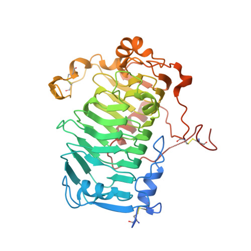
    6FI2

  • PubMed Abstract: 

    Capsules are surface layers of hydrated capsular polysaccharides (CPSs) produced by many bacteria. The human pathogen Salmonella enterica serovar Typhi produces "Vi antigen" CPS, which contributes to virulence. In a conserved strategy used by bacteria with diverse CPS structures, translocation of Vi antigen to the cell surface is driven by an ATP-binding cassette (ABC) transporter. These transporters are engaged in heterooligomeric complexes proposed to form an enclosed translocation conduit to the cell surface, allowing the transporter to power the entire process. We identified Vi antigen biosynthesis genetic loci in genera of the Burkholderiales , which are paradoxically distinguished from S. Typhi by encoding VexL, a predicted pectate lyase homolog. Biochemical analyses demonstrated that VexL is an unusual metal-independent endolyase with an acidic pH optimum that is specific for O-acetylated Vi antigen. A 1.22-Å crystal structure of the VexL-Vi antigen complex revealed features which distinguish common secreted catabolic pectate lyases from periplasmic VexL, which participates in cell-surface assembly. VexL possesses a right-handed parallel β-superhelix, of which one face forms an electropositive glycan-binding groove with an extensive hydrogen bonding network that includes Vi antigen acetyl groups and confers substrate specificity. VexL provided a probe to interrogate conserved features of the ABC transporter-dependent export model. When introduced into S Typhi, VexL localized to the periplasm and degraded Vi antigen. In contrast, a cytosolic derivative had no effect unless export was disrupted. These data provide evidence that CPS assembled in ABC transporter-dependent systems is actually exposed to the periplasm during envelope translocation.


  • Organizational Affiliation
    • Department of Molecular and Cellular Biology, University of Guelph, Guelph, ON N1G 2W1, Canada.

Macromolecule Content 

  • Total Structure Weight: 46.22 kDa 
  • Atom Count: 3,280 
  • Modeled Residue Count: 375 
  • Deposited Residue Count: 408 
  • Unique protein chains: 1

Macromolecules

Find similar proteins by:|  3D Structure
Entity ID: 1
MoleculeChains  Sequence LengthOrganismDetailsImage
VexL408Achromobacter denitrificansMutation(s): 0 
Gene Names: vexL
EC: 4.2.2.2
UniProt
Find proteins for A0A160EBC2 (Achromobacter denitrificans)
Explore A0A160EBC2 
Go to UniProtKB:  A0A160EBC2
Entity Groups
Sequence Clusters30% Identity50% Identity70% Identity90% Identity95% Identity100% Identity
UniProt GroupA0A160EBC2
Sequence Annotations
Expand
Reference Sequence

Oligosaccharides

Help  
Entity ID: 2
MoleculeChains Length2D Diagram GlycosylationD Interactions
2-acetamido-2-deoxy-alpha-D-galactopyranuronic acid-(1-4)-3-O-acetyl-2-acetamido-2-deoxy-alpha-D-galactopyranuronic acid-(1-4)-3-O-acetyl-2-acetamido-2-deoxy-alpha-D-galactopyranuronic acid
B
3N/A
Glycosylation Resources
GlyTouCan: G87382CT
GlyCosmos: G87382CT

Small Molecules

Ligands 1 Unique
IDChains Name / Formula / InChI Key2D Diagram3D Interactions
MLI

Query on MLI


Download:Ideal Coordinates CCD File
C [auth A]MALONATE ION
C3 H2 O4
OFOBLEOULBTSOW-UHFFFAOYSA-L

Experimental Data & Validation

Experimental Data

  • Method: X-RAY DIFFRACTION
  • Resolution: 1.22 Å
  • R-Value Free:  0.166 (Depositor), 0.166 (DCC) 
  • R-Value Work:  0.139 (Depositor), 0.139 (DCC) 
  • R-Value Observed: 0.140 (Depositor) 
Space Group: P 42 21 2
Unit Cell:
Length ( Å )Angle ( ˚ )
a = 94.68α = 90
b = 94.68β = 90
c = 78.84γ = 90
Software Package:
Software NamePurpose
REFMACrefinement
autoPROCdata scaling
BALBESphasing
XDSdata reduction

Structure Validation

View Full Validation Report


Entry History 

& Funding Information

Deposition Data


Funding OrganizationLocationGrant Number
Canada--

Revision History  (Full details and data files)

  • Version 1.0: 2018-05-02
    Type: Initial release
  • Version 1.1: 2018-05-30
    Changes: Data collection, Database references
  • Version 2.0: 2020-07-29
    Type: Remediation
    Reason: Carbohydrate remediation
    Changes: Atomic model, Data collection, Derived calculations, Refinement description, Structure summary
  • Version 2.1: 2024-11-06
    Changes: Data collection, Database references, Structure summary