6FH8

E. coli surface display of streptavidin for directed evolution of an allylic deallocase


Experimental Data Snapshot

  • Method: X-RAY DIFFRACTION
  • Resolution: 1.64 Å
  • R-Value Free: 0.159 
  • R-Value Work: 0.120 
  • R-Value Observed: 0.122 

wwPDB Validation   3D Report Full Report


Ligand Structure Quality Assessment 


This is version 1.0 of the entry. See complete history


Literature

E. colisurface display of streptavidin for directed evolution of an allylic deallylase.

Heinisch, T.Schwizer, F.Garabedian, B.Csibra, E.Jeschek, M.Vallapurackal, J.Pinheiro, V.B.Marliere, P.Panke, S.Ward, T.R.

(2018) Chem Sci 9: 5383-5388

  • DOI: https://doi.org/10.1039/c8sc00484f
  • Primary Citation of Related Structures:  
    6FH8

  • PubMed Abstract: 

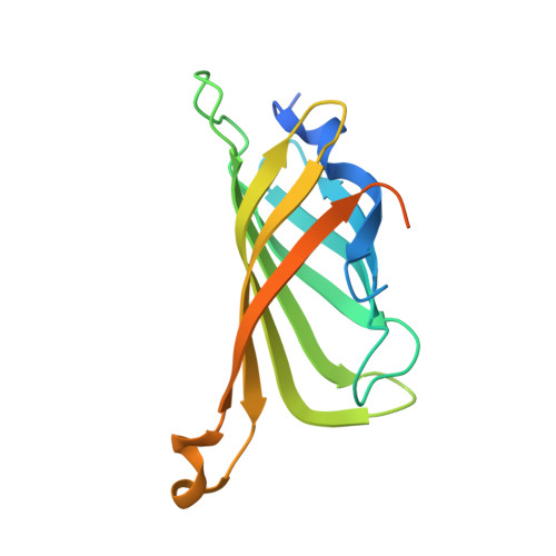
    Artificial metalloenzymes (ArMs hereafter) combine attractive features of both homogeneous catalysts and enzymes and offer the potential to implement new-to-nature reactions in living organisms. Herein we present an E. coli surface display platform for streptavidin (Sav hereafter) relying on an Lpp-OmpA anchor. The system was used for the high throughput screening of a bioorthogonal CpRu-based artificial deallylase (ADAse) that uncages an allylcarbamate-protected aminocoumarin 1 . Two rounds of directed evolution afforded the double mutant S112M-K121A that displayed a 36-fold increase in surface activity vs. cellular background and a 5.7-fold increased in vitro activity compared to the wild type enzyme. The crystal structure of the best ADAse reveals the importance of mutation S112M to stabilize the cofactor conformation inside the protein.


  • Organizational Affiliation

    Department of Chemistry , University of Basel , Mattenstrasse 24a , Basel CH-4002 , Switzerland . Email: thomas.ward@unibas.ch.


Macromolecules
Find similar proteins by:  (by identity cutoff)  |  3D Structure
Entity ID: 1
MoleculeChains Sequence LengthOrganismDetailsImage
Streptavidin159Streptomyces avidiniiMutation(s): 2 
UniProt
Find proteins for P22629 (Streptomyces avidinii)
Explore P22629 
Go to UniProtKB:  P22629
Entity Groups  
Sequence Clusters30% Identity50% Identity70% Identity90% Identity95% Identity100% Identity
UniProt GroupP22629
Sequence Annotations
Expand
  • Reference Sequence
Small Molecules
Ligands 1 Unique
IDChains Name / Formula / InChI Key2D Diagram3D Interactions
JCT
Query on JCT

Download Ideal Coordinates CCD File 
B [auth A]biotinylated ruthenium cyclopentadienide
C29 H28 N5 O5 Ru S
CQAGRAKHOFYKGA-XRCFJZOASA-L
Experimental Data & Validation

Experimental Data

  • Method: X-RAY DIFFRACTION
  • Resolution: 1.64 Å
  • R-Value Free: 0.159 
  • R-Value Work: 0.120 
  • R-Value Observed: 0.122 
  • Space Group: I 41 2 2
Unit Cell:
Length ( Å )Angle ( ˚ )
a = 57.722α = 90
b = 57.722β = 90
c = 183.615γ = 90
Software Package:
Software NamePurpose
REFMACrefinement
XDSdata reduction
Aimlessdata scaling
REFMACphasing

Structure Validation

View Full Validation Report


Ligand Structure Quality Assessment 


Entry History 

Deposition Data

Revision History  (Full details and data files)

  • Version 1.0: 2018-08-22
    Type: Initial release