6FGK | pdb_00006fgk

Crystal structure of the small alarmone synthethase 2 from Bacillus subtilis


Experimental Data Snapshot

  • Method: X-RAY DIFFRACTION
  • Resolution: 3.20 Å
  • R-Value Free: 
    0.319 (Depositor), 0.314 (DCC) 
  • R-Value Work: 
    0.257 (Depositor), 0.258 (DCC) 
  • R-Value Observed: 
    0.260 (Depositor) 

Starting Model: experimental
View more details

wwPDB Validation 3D Report Full Report

Validation slider image for 6FGK

This is version 1.2 of the entry. See complete history

Literature

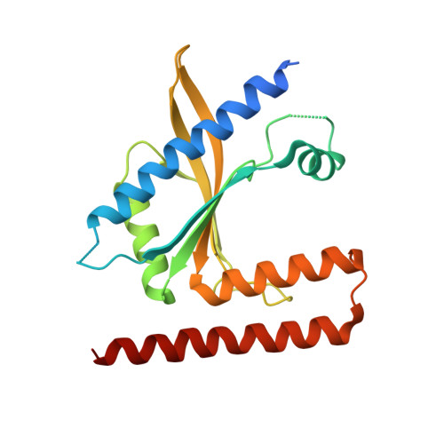
Structural and mechanistic divergence of the small (p)ppGpp synthetases RelP and RelQ.

Steinchen, W.Vogt, M.S.Altegoer, F.Giammarinaro, P.I.Horvatek, P.Wolz, C.Bange, G.

(2018) Sci Rep 8: 2195-2195

  • DOI: https://doi.org/10.1038/s41598-018-20634-4
  • Primary Citation Related Structures: 
    6FGJ, 6FGK, 6FGX

  • PubMed Abstract: 

    The nutritional alarmones ppGpp and pppGpp (collectively: (p)ppGpp) are nucleotide-based second messengers enabling bacteria to respond to environmental and stress conditions. Several bacterial species contain two highly homologous (p)ppGpp synthetases named RelP (SAS2, YwaC) and RelQ (SAS1, YjbM). It is established that RelQ forms homotetramers that are subject to positive allosteric regulation by pppGpp, but structural and mechanistic insights into RelP lack behind. Here we present a structural and mechanistic characterization of RelP. In stark contrast to RelQ, RelP is not allosterically regulated by pppGpp and displays a different enzyme kinetic behavior. This discrepancy is evoked by different conformational properties of the guanosine-substrate binding site (G-Loop) of both proteins. Our study shows how minor structural divergences between close homologues result in new functional features during the course of molecular evolution.


  • Organizational Affiliation
    • Philipps-University Marburg, LOEWE Center for Synthetic Microbiology & Department of Chemistry, Hans-Meerwein-Straße, 35043 Marburg, Germany. wieland.steinchen@synmikro.uni-marburg.de.

Macromolecule Content 

  • Total Structure Weight: 98.55 kDa 
  • Atom Count: 6,058 
  • Modeled Residue Count: 734 
  • Deposited Residue Count: 840 
  • Unique protein chains: 1

Macromolecules

Find similar proteins by:|  3D Structure
Entity ID: 1
MoleculeChains  Sequence LengthOrganismDetailsImage
GTP pyrophosphokinase YwaC
A, B, C, D
210Bacillus subtilis subsp. subtilis str. 168Mutation(s): 0 
Gene Names: ywaCBSU38480ipa-7d
EC: 2.7.6.5
UniProt
Find proteins for P39583 (Bacillus subtilis (strain 168))
Explore P39583 
Go to UniProtKB:  P39583
Entity Groups
Sequence Clusters30% Identity50% Identity70% Identity90% Identity95% Identity100% Identity
UniProt GroupP39583
Sequence Annotations
Expand
Reference Sequence

Experimental Data & Validation

Experimental Data

  • Method: X-RAY DIFFRACTION
  • Resolution: 3.20 Å
  • R-Value Free:  0.319 (Depositor), 0.314 (DCC) 
  • R-Value Work:  0.257 (Depositor), 0.258 (DCC) 
  • R-Value Observed: 0.260 (Depositor) 
Space Group: P 21 21 21
Unit Cell:
Length ( Å )Angle ( ˚ )
a = 83.67α = 90
b = 99.92β = 90
c = 122.884γ = 90
Software Package:
Software NamePurpose
PHENIXrefinement
XDSdata reduction
Aimlessdata scaling
PHASERphasing

Structure Validation

View Full Validation Report


Entry History 

Deposition Data

Revision History  (Full details and data files)

  • Version 1.0: 2018-02-07
    Type: Initial release
  • Version 1.1: 2018-02-14
    Changes: Database references
  • Version 1.2: 2024-01-17
    Changes: Data collection, Database references, Refinement description