6FGE

Crystal structure of human ZUFSP/ZUP1 in complex with ubiquitin


Experimental Data Snapshot

  • Method: X-RAY DIFFRACTION
  • Resolution: 1.74 Å
  • R-Value Free: 0.209 
  • R-Value Work: 0.174 
  • R-Value Observed: 0.175 

wwPDB Validation   3D Report Full Report


This is version 1.2 of the entry. See complete history


Literature

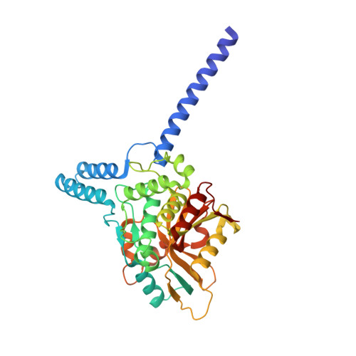
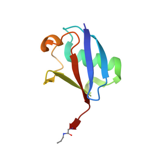
Discovery and Characterization of ZUFSP/ZUP1, a Distinct Deubiquitinase Class Important for Genome Stability.

Kwasna, D.Abdul Rehman, S.A.Natarajan, J.Matthews, S.Madden, R.De Cesare, V.Weidlich, S.Virdee, S.Ahel, I.Gibbs-Seymour, I.Kulathu, Y.

(2018) Mol Cell 70: 150-164.e6

  • DOI: https://doi.org/10.1016/j.molcel.2018.02.023
  • Primary Citation of Related Structures:  
    6FGE

  • PubMed Abstract: 
  • Deubiquitinating enzymes (DUBs) are important regulators of ubiquitin signaling. Here, we report the discovery of deubiquitinating activity in ZUFSP/C6orf113. High-resolution crystal structures of ZUFSP in complex with ubiquitin reveal several distinctive features of ubiquitin recognition and catalysis ...

    Deubiquitinating enzymes (DUBs) are important regulators of ubiquitin signaling. Here, we report the discovery of deubiquitinating activity in ZUFSP/C6orf113. High-resolution crystal structures of ZUFSP in complex with ubiquitin reveal several distinctive features of ubiquitin recognition and catalysis. Our analyses reveal that ZUFSP is a novel DUB with no homology to any known DUBs, leading us to classify ZUFSP as the seventh DUB family. Intriguingly, the minimal catalytic domain does not cleave polyubiquitin. We identify two ubiquitin binding domains in ZUFSP: a ZHA (ZUFSP helical arm) that binds to the distal ubiquitin and an atypical UBZ domain in ZUFSP that binds to polyubiquitin. Importantly, both domains are essential for ZUFSP to selectively cleave K63-linked polyubiquitin. We show that ZUFSP localizes to DNA lesions, where it plays an important role in genome stability pathways, functioning to prevent spontaneous DNA damage and also promote cellular survival in response to exogenous DNA damage.


    Organizational Affiliation

    MRC Protein Phosphorylation & Ubiquitylation Unit, School of Life Sciences, University of Dundee, Dow Street, Dundee DD1 5EH, UK. Electronic address: ykulathu@dundee.ac.uk.


Macromolecules
Find similar proteins by:  (by identity cutoff)  |  3D Structure
Entity ID: 1
MoleculeChainsSequence LengthOrganismDetailsImage
Zinc finger with UFM1-specific peptidase domain protein348Homo sapiensMutation(s): 0 
Gene Names: ZUFSPC6orf113ZUP1
EC: 3.4.19.12
UniProt & NIH Common Fund Data Resources
Find proteins for Q96AP4 (Homo sapiens)
Explore Q96AP4 
Go to UniProtKB:  Q96AP4
PHAROS:  Q96AP4
Entity Groups  
Sequence Clusters30% Identity50% Identity70% Identity90% Identity95% Identity100% Identity
UniProt GroupQ96AP4
Protein Feature View
Expand
  • Reference Sequence
Find similar proteins by:  (by identity cutoff)  |  3D Structure
Entity ID: 2
MoleculeChainsSequence LengthOrganismDetailsImage
Polyubiquitin-BB [auth C]76Homo sapiensMutation(s): 1 
Gene Names: UBB
UniProt & NIH Common Fund Data Resources
Find proteins for P0CG47 (Homo sapiens)
Explore P0CG47 
Go to UniProtKB:  P0CG47
PHAROS:  P0CG47
Entity Groups  
Sequence Clusters30% Identity50% Identity70% Identity90% Identity95% Identity100% Identity
UniProt GroupP0CG47
Protein Feature View
Expand
  • Reference Sequence
Small Molecules
Ligands 7 Unique
IDChainsName / Formula / InChI Key2D Diagram3D Interactions
PEG
Query on PEG

Download Ideal Coordinates CCD File 
O [auth A]DI(HYDROXYETHYL)ETHER
C4 H10 O3
MTHSVFCYNBDYFN-UHFFFAOYSA-N
 Ligand Interaction
MLI
Query on MLI

Download Ideal Coordinates CCD File 
C [auth A]MALONATE ION
C3 H2 O4
OFOBLEOULBTSOW-UHFFFAOYSA-L
 Ligand Interaction
GOL
Query on GOL

Download Ideal Coordinates CCD File 
G [auth A],
H [auth A]
GLYCEROL
C3 H8 O3
PEDCQBHIVMGVHV-UHFFFAOYSA-N
 Ligand Interaction
EDO
Query on EDO

Download Ideal Coordinates CCD File 
D [auth A],
E [auth A],
F [auth A]
1,2-ETHANEDIOL
C2 H6 O2
LYCAIKOWRPUZTN-UHFFFAOYSA-N
 Ligand Interaction
ACT
Query on ACT

Download Ideal Coordinates CCD File 
R [auth C]ACETATE ION
C2 H3 O2
QTBSBXVTEAMEQO-UHFFFAOYSA-M
 Ligand Interaction
FMT
Query on FMT

Download Ideal Coordinates CCD File 
I [auth A]
J [auth A]
K [auth A]
L [auth A]
M [auth A]
I [auth A],
J [auth A],
K [auth A],
L [auth A],
M [auth A],
N [auth A],
S [auth C]
FORMIC ACID
C H2 O2
BDAGIHXWWSANSR-UHFFFAOYSA-N
 Ligand Interaction
NH4
Query on NH4

Download Ideal Coordinates CCD File 
P [auth A],
Q [auth A]
AMMONIUM ION
H4 N
QGZKDVFQNNGYKY-UHFFFAOYSA-O
 Ligand Interaction
Experimental Data & Validation

Experimental Data

  • Method: X-RAY DIFFRACTION
  • Resolution: 1.74 Å
  • R-Value Free: 0.209 
  • R-Value Work: 0.174 
  • R-Value Observed: 0.175 
  • Space Group: P 65 2 2
Unit Cell:
Length ( Å )Angle ( ˚ )
a = 84.484α = 90
b = 84.484β = 90
c = 201.867γ = 120
Software Package:
Software NamePurpose
XDSdata reduction
XDSdata scaling
REFMACrefinement

Structure Validation

View Full Validation Report


Entry History & Funding Information

Deposition Data


Funding OrganizationLocationGrant Number
Medical Research Council (United Kingdom)United KingdomMC_UU_12016/6
European Research CouncilUnited Kingdom677623

Revision History  (Full details and data files)

  • Version 1.0: 2018-04-04
    Type: Initial release
  • Version 1.1: 2018-04-11
    Changes: Data collection, Derived calculations, Structure summary
  • Version 1.2: 2018-04-18
    Changes: Data collection, Database references