Directed Evolution of Alcohol Dehydrogenase for Improved Stereoselective Redox Transformations of 1-Phenylethane-1,2-diol and Its Corresponding Acyloin.
Hamnevik, E., Maurer, D., Enugala, T.R., Chu, T., Lofgren, R., Dobritzsch, D., Widersten, M.(2018) Biochemistry 57: 1059-1062
- PubMed: 29384657 Search on PubMed
- DOI: https://doi.org/10.1021/acs.biochem.8b00055
- Primary Citation Related Structures:
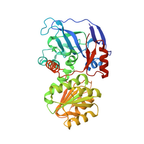
6FFX, 6FFZ - PubMed Abstract:
Laboratory evolution of alcohol dehydrogenase produced enzyme variants with improved turnover numbers with a vicinal 1,2-diol and its corresponding hydroxyketone. Crystal structure and transient kinetics analysis aids in rationalizing the new functions of these variants.
- Department of Chemistry-BMC, Uppsala University , Box 576, SE-751 23 Uppsala, Sweden.
Organizational Affiliation: