6FDF | pdb_00006fdf

Crystal structure of S. pombe Dnmt2 methyltransferase


Experimental Data Snapshot

  • Method: X-RAY DIFFRACTION
  • Resolution: 1.70 Å
  • R-Value Free: 
    0.195 (Depositor), 0.198 (DCC) 
  • R-Value Work: 
    0.174 (Depositor), 0.177 (DCC) 
  • R-Value Observed: 
    0.174 (Depositor) 

wwPDB Validation 3D Report Full Report

Validation slider image for 6FDF

Ligand Structure Quality Assessment 


This is version 1.2 of the entry. See complete history

Literature

Structural insights into the stimulation of S. pombe Dnmt2 catalytic efficiency by the tRNA nucleoside queuosine.

Johannsson, S.Neumann, P.Wulf, A.Welp, L.M.Gerber, H.D.Krull, M.Diederichsen, U.Urlaub, H.Ficner, R.

(2018) Sci Rep 8: 8880-8880

  • DOI: https://doi.org/10.1038/s41598-018-27118-5
  • Primary Citation Related Structures: 
    6FDF

  • PubMed Abstract: 

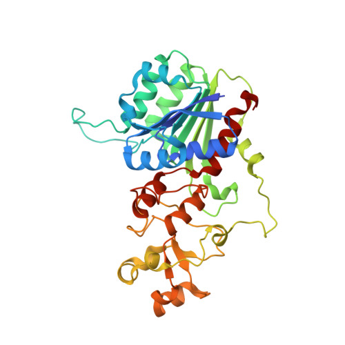
    Dnmt2 methylates cytosine at position 38 of tRNA Asp in a variety of eukaryotic organisms. A correlation between the presence of the hypermodified nucleoside queuosine (Q) at position 34 of tRNA Asp and the Dnmt2 dependent C38 methylation was recently found in vivo for S. pombe and D. discoideum. We demonstrate a direct effect of the Q-modification on the methyltransferase catalytic efficiency in vitro, as V max /K 0.5 of purified S. pombe Dnmt2 shows an increase for in vitro transcribed tRNA Asp containing Q34 to 6.27 ∗ 10 -3  s -1  µM -1 compared to 1.51 ∗ 10 -3  s -1  µM -1 for the unmodified substrate. Q34tRNA Asp exhibits an only slightly increased affinity for Dnmt2 in comparison to unmodified G34tRNA. In order to get insight into the structural basis for the Q-dependency, the crystal structure of S. pombe Dnmt2 was determined at 1.7 Å resolution. It closely resembles the known structures of human and E. histolytica Dnmt2, and contains the entire active site loop. The interaction with tRNA was analyzed by means of mass-spectrometry using UV cross-linked Dnmt2-tRNA complex. These cross-link data and computational docking of Dnmt2 and tRNA Asp reveal Q34 positioned adjacent to the S-adenosylmethionine occupying the active site, suggesting that the observed increase of Dnmt2 catalytic efficiency by queuine originates from optimal positioning of the substrate molecules and residues relevant for methyl transfer.


  • Organizational Affiliation
    • Department of Molecular Structural Biology, Institute of Microbiology and Genetics, GZMB, Georg-August-University Göttingen, 37077, Göttingen, Germany.

Macromolecule Content 

  • Total Structure Weight: 155.75 kDa 
  • Atom Count: 11,492 
  • Modeled Residue Count: 1,269 
  • Deposited Residue Count: 1,336 
  • Unique protein chains: 1

Macromolecules

Find similar proteins by:|  3D Structure
Entity ID: 1
MoleculeChains  Sequence LengthOrganismDetailsImage
tRNA (cytosine(38)-C(5))-methyltransferase
A, B, C, D
334Schizosaccharomyces pombeMutation(s): 0 
Gene Names: pmt1SPBC19C2.02
EC: 2.1.1.204
UniProt
Find proteins for P40999 (Schizosaccharomyces pombe (strain 972 / ATCC 24843))
Explore P40999 
Go to UniProtKB:  P40999
Entity Groups
Sequence Clusters30% Identity50% Identity70% Identity90% Identity95% Identity100% Identity
UniProt GroupP40999
Sequence Annotations
Expand
Reference Sequence

Small Molecules

Ligands 2 Unique
IDChains Name / Formula / InChI Key2D Diagram3D Interactions
SAH

Query on SAH


Download:Ideal Coordinates CCD File
E [auth A],
J [auth B],
N [auth C],
Q [auth D]
S-ADENOSYL-L-HOMOCYSTEINE
C14 H20 N6 O5 S
ZJUKTBDSGOFHSH-WFMPWKQPSA-N
GOL

Query on GOL


Download:Ideal Coordinates CCD File
F [auth A]
G [auth A]
H [auth A]
I [auth A]
K [auth B]
F [auth A],
G [auth A],
H [auth A],
I [auth A],
K [auth B],
L [auth B],
M [auth B],
O [auth C],
P [auth C],
R [auth D],
S [auth D]
GLYCEROL
C3 H8 O3
PEDCQBHIVMGVHV-UHFFFAOYSA-N

Experimental Data & Validation

Experimental Data

  • Method: X-RAY DIFFRACTION
  • Resolution: 1.70 Å
  • R-Value Free:  0.195 (Depositor), 0.198 (DCC) 
  • R-Value Work:  0.174 (Depositor), 0.177 (DCC) 
  • R-Value Observed: 0.174 (Depositor) 
Space Group: C 1 2 1
Unit Cell:
Length ( Å )Angle ( ˚ )
a = 153.817α = 90
b = 114.781β = 131.05
c = 113.759γ = 90
Software Package:
Software NamePurpose
XDSdata reduction
XSCALEdata scaling
PHENIXrefinement
PDB_EXTRACTdata extraction
PHASERphasing

Structure Validation

View Full Validation Report


Ligand Structure Quality Assessment 


Entry History 

& Funding Information

Deposition Data


Funding OrganizationLocationGrant Number
German Research FoundationGermanySPP1784

Revision History  (Full details and data files)

  • Version 1.0: 2018-06-20
    Type: Initial release
  • Version 1.1: 2024-05-08
    Changes: Data collection, Database references
  • Version 1.2: 2025-12-10
    Changes: Data collection, Structure summary