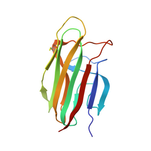
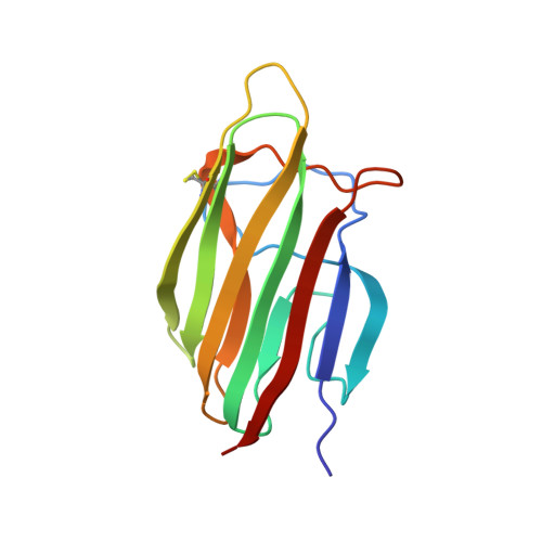
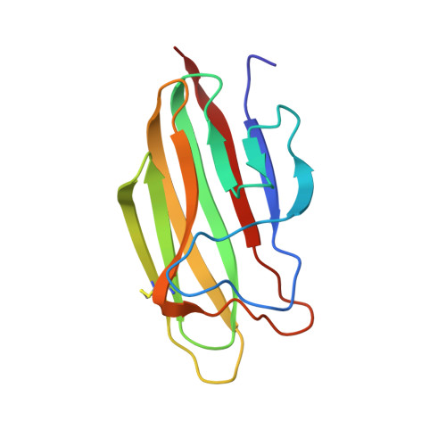
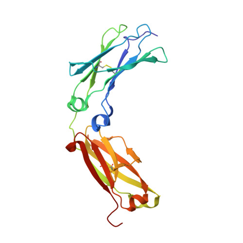
Structures of C1-IgG1 provide insights into how danger pattern recognition activates complement.
Ugurlar, D., Howes, S.C., de Kreuk, B.J., Koning, R.I., de Jong, R.N., Beurskens, F.J., Schuurman, J., Koster, A.J., Sharp, T.H., Parren, P.W.H.I., Gros, P.(2018) Science 359: 794-797
- PubMed: 29449492
- DOI: https://doi.org/10.1126/science.aao4988
- Primary Citation of Related Structures:
6FCZ - PubMed Abstract:
Danger patterns on microbes or damaged host cells bind and activate C1, inducing innate immune responses and clearance through the complement cascade. How these patterns trigger complement initiation remains elusive. Here, we present cryo-electron microscopy analyses of C1 bound to monoclonal antibodies in which we observed heterogeneous structures of single and clustered C1-immunoglobulin G1 (IgG1) hexamer complexes. Distinct C1q binding sites are observed on the two Fc-CH2 domains of each IgG molecule. These are consistent with known interactions and also reveal additional interactions, which are supported by functional IgG1-mutant analysis. Upon antibody binding, the C1q arms condense, inducing rearrangements of the C1r 2 s 2 proteases and tilting C1q's cone-shaped stalk. The data suggest that C1r may activate C1s within single, strained C1 complexes or between neighboring C1 complexes on surfaces.
- Crystal and Structural Chemistry, Bijvoet Center for Biomolecular Research, Department of Chemistry, Faculty of Science, Utrecht University, Padualaan 8, 3584 CH Utrecht, Netherlands.
Organizational Affiliation:


