6FBD | pdb_00006fbd

KlenTaq DNA polymerase processing a modified primer - bearing the modification upstream at the second primer nucleotide.


Experimental Data Snapshot

  • Method: X-RAY DIFFRACTION
  • Resolution: 2.10 Å
  • R-Value Free: 
    0.233 (Depositor), 0.236 (DCC) 
  • R-Value Work: 
    0.198 (Depositor), 0.200 (DCC) 
  • R-Value Observed: 
    0.200 (Depositor) 

wwPDB Validation 3D Report Full Report

Validation slider image for 6FBD

Ligand Structure Quality Assessment 


This is version 1.3 of the entry. See complete history

Literature

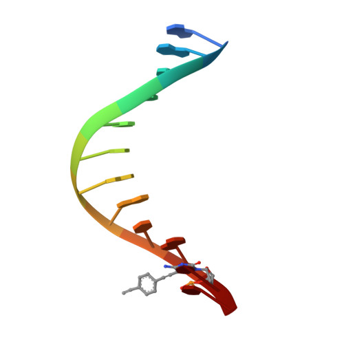
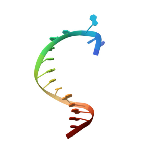
Snapshots of a modified nucleotide moving through the confines of a DNA polymerase.

Kropp, H.M.Durr, S.L.Peter, C.Diederichs, K.Marx, A.

(2018) Proc Natl Acad Sci U S A 115: 9992-9997

  • DOI: https://doi.org/10.1073/pnas.1811518115
  • Primary Citation Related Structures: 
    6FBC, 6FBD, 6FBE, 6FBF, 6FBG, 6FBH, 6FBI

  • PubMed Abstract: 

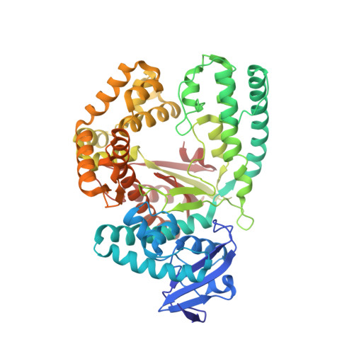
    DNA polymerases have evolved to process the four canonical nucleotides accurately. Nevertheless, these enzymes are also known to process modified nucleotides, which is the key to numerous core biotechnology applications. Processing of modified nucleotides includes incorporation of the modified nucleotide and postincorporation elongation to proceed with the synthesis of the nascent DNA strand. The structural basis for postincorporation elongation is currently unknown. We addressed this issue and successfully crystallized KlenTaq DNA polymerase in six closed ternary complexes containing the enzyme, the modified DNA substrate, and the incoming nucleotide. Each structure shows a high-resolution snapshot of the elongation of a modified primer, where the modification "moves" from the 3'-primer terminus upstream to the sixth nucleotide in the primer strand. Combining these data with quantum mechanics/molecular mechanics calculations and biochemical studies elucidates how the enzyme and the modified substrate mutually modulate their conformations without compromising the enzyme's activity significantly. The study highlights the plasticity of the system as origin of the broad substrate properties of DNA polymerases and facilitates the design of improved systems.


  • Organizational Affiliation
    • Department of Chemistry, Universität Konstanz, 78464 Konstanz, Germany.

Macromolecule Content 

  • Total Structure Weight: 70.51 kDa 
  • Atom Count: 4,930 
  • Modeled Residue Count: 567 
  • Deposited Residue Count: 569 
  • Unique protein chains: 1
  • Unique nucleic acid chains: 2

Macromolecules


Find similar proteins by:|  3D Structure
Entity ID: 1
MoleculeChains  Sequence LengthOrganismDetailsImage
DNA polymerase I, thermostable541Thermus aquaticusMutation(s): 0 
Gene Names: polApol1
EC: 2.7.7.7
UniProt
Find proteins for P19821 (Thermus aquaticus)
Explore P19821 
Go to UniProtKB:  P19821
Entity Groups
Sequence Clusters30% Identity50% Identity70% Identity90% Identity95% Identity100% Identity
UniProt GroupP19821
Sequence Annotations
Expand
Reference Sequence
Find similar nucleic acids by:  Sequence
Entity ID: 2
MoleculeChains LengthOrganismImage
DNA (5'-D(*GP*AP*CP*CP*AP*CP*GP*GP*CP*AP*(OH3)P*C)-3')12synthetic construct
Sequence Annotations
Expand
Reference Sequence
Find similar nucleic acids by:  Sequence
Entity ID: 3
MoleculeChains LengthOrganismImage
DNA (5'-D(P*AP*AP*AP*CP*GP*GP*TP*GP*CP*CP*GP*TP*GP*GP*TP*C)-3')16synthetic construct
Sequence Annotations
Expand
Reference Sequence

Small Molecules

Ligands 4 Unique
IDChains Name / Formula / InChI Key2D Diagram3D Interactions
XG4

Query on XG4


Download:Ideal Coordinates CCD File
F [auth A]2'-deoxy-5'-O-[(R)-hydroxy{[(R)-hydroxy(phosphonooxy)phosphoryl]amino}phosphoryl]guanosine
C10 H17 N6 O12 P3
DWGAAFQEGIMTIA-KVQBGUIXSA-N
GOL

Query on GOL


Download:Ideal Coordinates CCD File
H [auth C]GLYCEROL
C3 H8 O3
PEDCQBHIVMGVHV-UHFFFAOYSA-N
MN

Query on MN


Download:Ideal Coordinates CCD File
D [auth A],
E [auth A]
MANGANESE (II) ION
Mn
WAEMQWOKJMHJLA-UHFFFAOYSA-N
MG

Query on MG


Download:Ideal Coordinates CCD File
G [auth B],
I [auth C]
MAGNESIUM ION
Mg
JLVVSXFLKOJNIY-UHFFFAOYSA-N

Experimental Data & Validation

Experimental Data

  • Method: X-RAY DIFFRACTION
  • Resolution: 2.10 Å
  • R-Value Free:  0.233 (Depositor), 0.236 (DCC) 
  • R-Value Work:  0.198 (Depositor), 0.200 (DCC) 
  • R-Value Observed: 0.200 (Depositor) 
Space Group: P 31 2 1
Unit Cell:
Length ( Å )Angle ( ˚ )
a = 109.964α = 90
b = 109.964β = 90
c = 91.165γ = 120
Software Package:
Software NamePurpose
PHENIXrefinement
XDSdata reduction
XDSdata scaling
PHENIXphasing

Structure Validation

View Full Validation Report


Ligand Structure Quality Assessment 


Entry History 

& Funding Information

Deposition Data


Funding OrganizationLocationGrant Number
German Research FoundationGermany--

Revision History  (Full details and data files)

  • Version 1.0: 2018-09-26
    Type: Initial release
  • Version 1.1: 2018-10-10
    Changes: Data collection, Database references, Structure summary
  • Version 1.2: 2019-08-14
    Changes: Data collection, Structure summary
  • Version 1.3: 2024-05-08
    Changes: Data collection, Database references, Derived calculations