6FAN | pdb_00006fan

Crystal structure of putative CooT from Carboxydothermus hydrogenoformans


Experimental Data Snapshot

  • Method: X-RAY DIFFRACTION
  • Resolution: 2.00 Å
  • R-Value Free: 
    0.275 (Depositor), 0.237 (DCC) 
  • R-Value Work: 
    0.235 (Depositor), 0.238 (DCC) 
  • R-Value Observed: 
    0.237 (Depositor) 

wwPDB Validation 3D Report Full Report

Validation slider image for 6FAN

This is version 1.2 of the entry. See complete history

Literature

Biophysical and structural characterization of the putative nickel chaperone CooT from Carboxydothermus hydrogenoformans.

Alfano, M.Perard, J.Miras, R.Catty, P.Cavazza, C.

(2018) J Biol Inorg Chem 23: 809-817

  • DOI: https://doi.org/10.1007/s00775-018-1576-2
  • Primary Citation Related Structures: 
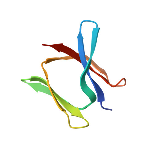
    6FAN

  • PubMed Abstract: 

    Carboxydothermus hydrogenoformans is a model microorganism for the study of [NiFe]-CODH, a key enzyme of carbon cycle in anaerobic microorganisms. The enzyme possesses a unique active site (C-cluster), constituted of a distorted [NiFe 3 S 4 ] cubane linked to a mononuclear Fe(II) center. Both the biogenesis of the C-cluster and the activation of CODH by nickel insertion remain unclear. Among the three accessory proteins thought to play a role in this latter step (CooC, CooJ, and CooT), CooT is identified as a nickel chaperone involved in CODH maturation in Rhodospirillum rubrum. Here, we structurally and biophysically characterized a putative CooT protein present in C. hydrogenoformans (pChCooT). Despite the low sequence homologies between CooT from R. rubrum (RrCooT) and pChCooT (19% sequence identity), the two proteins share several similarities, such as their overall structure and a solvent-exposed Ni(II)-binding site at the dimer interface. Moreover, the X-ray structure of pChCooT reveals the proximity between the histidine 55, a potential nickel-coordinating residue, and the cysteine 2, a highly conserved key residue in Ni(II)-binding.


  • Organizational Affiliation
    • University of Grenoble Alpes, CEA, CNRS, BIG, CBM, 38000, Grenoble, France.

Macromolecule Content 

  • Total Structure Weight: 44.66 kDa 
  • Atom Count: 3,093 
  • Modeled Residue Count: 368 
  • Deposited Residue Count: 378 
  • Unique protein chains: 1

Macromolecules

Find similar proteins by:|  3D Structure
Entity ID: 1
MoleculeChains  Sequence LengthOrganismDetailsImage
CooT
A, B, C, D, E
A, B, C, D, E, F
63Carboxydothermus hydrogenoformans Z-2901Mutation(s): 0 
Gene Names: CHY_0178
UniProt
Find proteins for Q3AFN4 (Carboxydothermus hydrogenoformans (strain ATCC BAA-161 / DSM 6008 / Z-2901))
Explore Q3AFN4 
Go to UniProtKB:  Q3AFN4
Entity Groups
Sequence Clusters30% Identity50% Identity70% Identity90% Identity95% Identity100% Identity
UniProt GroupQ3AFN4
Sequence Annotations
Expand
Reference Sequence

Small Molecules

Ligands 2 Unique
IDChains Name / Formula / InChI Key2D Diagram3D Interactions
MPD

Query on MPD


Download:Ideal Coordinates CCD File
G [auth B],
J [auth C],
K [auth D],
L [auth E]
(4S)-2-METHYL-2,4-PENTANEDIOL
C6 H14 O2
SVTBMSDMJJWYQN-YFKPBYRVSA-N
GOL

Query on GOL


Download:Ideal Coordinates CCD File
H [auth B],
I [auth B],
M [auth E],
N [auth E]
GLYCEROL
C3 H8 O3
PEDCQBHIVMGVHV-UHFFFAOYSA-N

Experimental Data & Validation

Experimental Data

  • Method: X-RAY DIFFRACTION
  • Resolution: 2.00 Å
  • R-Value Free:  0.275 (Depositor), 0.237 (DCC) 
  • R-Value Work:  0.235 (Depositor), 0.238 (DCC) 
  • R-Value Observed: 0.237 (Depositor) 
Space Group: P 1 21 1
Unit Cell:
Length ( Å )Angle ( ˚ )
a = 41.261α = 90
b = 61.35β = 100.41
c = 81.118γ = 90
Software Package:
Software NamePurpose
REFMACrefinement
XDSdata reduction
XSCALEdata scaling
PHASERphasing

Structure Validation

View Full Validation Report


Entry History 

Deposition Data

Revision History  (Full details and data files)

  • Version 1.0: 2018-11-28
    Type: Initial release
  • Version 1.1: 2019-03-27
    Changes: Data collection, Database references
  • Version 1.2: 2024-05-08
    Changes: Data collection, Database references