Inhibition of BUB1 Kinase by BAY 1816032 Sensitizes Tumor Cells toward Taxanes, ATR, and PARP InhibitorsIn VitroandIn Vivo.
Siemeister, G., Mengel, A., Fernandez-Montalvan, A.E., Bone, W., Schroder, J., Zitzmann-Kolbe, S., Briem, H., Prechtl, S., Holton, S.J., Monning, U., von Ahsen, O., Johanssen, S., Cleve, A., Putter, V., Hitchcock, M., von Nussbaum, F., Brands, M., Ziegelbauer, K., Mumberg, D.(2019) Clin Cancer Res 25: 1404-1414
- PubMed: 30429199 Search on PubMed
- DOI: https://doi.org/10.1158/1078-0432.CCR-18-0628
- Primary Citation Related Structures:
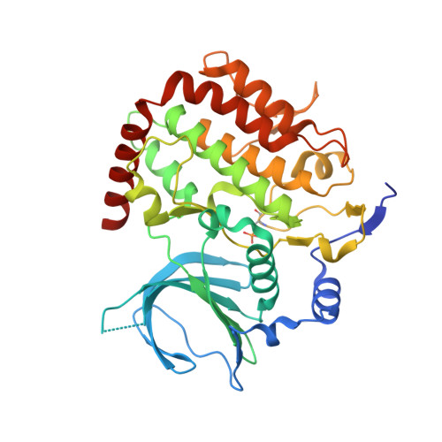
6F7B - PubMed Abstract:
The catalytic function of BUB1 is required for chromosome arm resolution and positioning of the chromosomal passenger complex for resolution of spindle attachment errors and plays only a minor role in spindle assembly checkpoint activation. Here, we present the identification and preclinical pharmacologic profile of the first BUB1 kinase inhibitor with good bioavailability. The Bayer compound library was screened for BUB1 kinase inhibitors and medicinal chemistry efforts to improve target affinity and physicochemical and pharmacokinetic parameters resulting in the identification of BAY 1816032 were performed. BAY 1816032 was characterized for kinase selectivity, inhibition of BUB1 signaling, and inhibition of tumor cell proliferation alone and in combination with taxanes, ATR, and PARP inhibitors. Effects on tumor growth in vivo were evaluated using human triple-negative breast xenograft models. The highly selective compound BAY 1816032 showed long target residence time and induced chromosome mis-segregation upon combination with low concentrations of paclitaxel. It was synergistic or additive in combination with paclitaxel or docetaxel, as well as with ATR or PARP inhibitors in cellular assays. Tumor xenograft studies demonstrated a strong and statistically significant reduction of tumor size and excellent tolerability upon combination of BAY 1816032 with paclitaxel or olaparib as compared with the respective monotherapies. Our findings suggest clinical proof-of-concept studies evaluating BAY 1816032 in combination with taxanes or PARP inhibitors to enhance their efficacy and potentially overcome resistance.
- Bayer AG, Muellerstrasse Berlin, Germany. gerhard.siemeister@bayer.com.
Organizational Affiliation: