6F6Z | pdb_00006f6z

Mouse Thymidylate Synthase Cocrystallized with N(4)OHdCMP and Soaked in Methylenetetrahydrofolate


Experimental Data Snapshot

  • Method: X-RAY DIFFRACTION
  • Resolution: 2.13 Å
  • R-Value Free: 
    0.243 (Depositor), 0.243 (DCC) 
  • R-Value Work: 
    0.210 (Depositor), 0.213 (DCC) 
  • R-Value Observed: 
    0.212 (Depositor) 

Starting Model: experimental
View more details

wwPDB Validation 3D Report Full Report

Validation slider image for 6F6Z

Ligand Structure Quality Assessment 


This is version 1.3 of the entry. See complete history

Literature

Molecular Mechanism of Thymidylate Synthase Inhibition by N4-Hydroxy-dCMP in View of Spectrophotometric and Crystallographic Studies

Maj, P.Jarmula, A.Wilk, P.Prokopowicz, M.Rypniewski, W.Zielinski, Z.Dowiercial, A.Bzowska, A.Rode, W.

(2021) Int J Mol Sci 22

  • DOI: https://doi.org/10.3390/ijms22094758
  • Primary Citation Related Structures: 
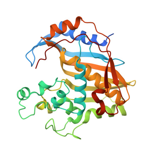
    5M4Z, 6F6Z

  • PubMed Abstract: 

    Novel evidence is presented allowing further clarification of the mechanism of the slow-binding thymidylate synthase (TS) inhibition by N 4 -hydroxy-dCMP (N 4 -OH-dCMP). Spectrophotometric monitoring documented time- and temperature-, and N 4 -OH-dCMP-dependent TS-catalyzed dihydrofolate production, accompanying the mouse enzyme incubation with N 4 -OH-dCMP and N 5,10 -methylenetetrahydrofolate, known to inactivate the enzyme by the covalent binding of the inhibitor, suggesting the demonstrated reaction to be uncoupled from the pyrimidine C(5) methylation. The latter was in accord with the hypothesis based on the previously presented structure of mouse TS (cf. PDB ID: 4EZ8), and with conclusions based on the present structure of the parasitic nematode Trichinella spiralis , both co-crystallized with N 4 -OH-dCMP and N 5,10 -methylenetetrahdrofolate. The crystal structure of the mouse TS-N 4 -OH-dCMP complex soaked with N 5,10 -methylenetetrahydrofolate revealed the reaction to run via a unique imidazolidine ring opening, leaving the one-carbon group bound to the N(10) atom, thus too distant from the pyrimidine C(5) atom to enable the electrophilic attack and methylene group transfer.


  • Organizational Affiliation
    • Nencki Institute of Experimental Biology, Polish Academy of Sciences, 3 Pasteur St., PL-02-093 Warszawa, Poland.

Macromolecule Content 

  • Total Structure Weight: 71.57 kDa 
  • Atom Count: 5,008 
  • Modeled Residue Count: 574 
  • Deposited Residue Count: 614 
  • Unique protein chains: 1

Macromolecules

Find similar proteins by:|  3D Structure
Entity ID: 1
MoleculeChains  Sequence LengthOrganismDetailsImage
Thymidylate synthase
A, B
307Mus musculusMutation(s): 0 
Gene Names: Tyms
EC: 2.1.1.45
UniProt
Find proteins for P07607 (Mus musculus)
Explore P07607 
Go to UniProtKB:  P07607
Entity Groups
Sequence Clusters30% Identity50% Identity70% Identity90% Identity95% Identity100% Identity
UniProt GroupP07607
Sequence Annotations
Expand
Reference Sequence

Small Molecules

Ligands 2 Unique
IDChains Name / Formula / InChI Key2D Diagram3D Interactions
TGQ

Query on TGQ


Download:Ideal Coordinates CCD File
D [auth A],
F [auth B]
(2~{S})-2-[[4-[[(6~{R})-2-azanyl-4-oxidanylidene-5,6,7,8-tetrahydro-1~{H}-pteridin-6-yl]methyl-methyl-amino]phenyl]carbonylamino]pentanedioic acid
C20 H25 N7 O6
ZHKQAQKGVKBJPP-YPMHNXCESA-N
NOH

Query on NOH


Download:Ideal Coordinates CCD File
C [auth A],
E [auth B]
2'-deoxy-N-hydroxycytidine 5'-(dihydrogen phosphate)
C9 H14 N3 O8 P
YKOWBJJOJNGCAD-SHYZEUOFSA-N
Binding Affinity Annotations 
IDSourceBinding Affinity
NOH BindingDB:  6F6Z Kd: 1000 (nM) from 1 assay(s)

Experimental Data & Validation

Experimental Data

  • Method: X-RAY DIFFRACTION
  • Resolution: 2.13 Å
  • R-Value Free:  0.243 (Depositor), 0.243 (DCC) 
  • R-Value Work:  0.210 (Depositor), 0.213 (DCC) 
  • R-Value Observed: 0.212 (Depositor) 
Space Group: C 1 2 1
Unit Cell:
Length ( Å )Angle ( ˚ )
a = 160.36α = 90
b = 89.16β = 97.47
c = 66.56γ = 90
Software Package:
Software NamePurpose
PHENIXrefinement
DENZOdata reduction
SCALEPACKdata scaling
PHASERphasing

Structure Validation

View Full Validation Report


Ligand Structure Quality Assessment 


Entry History 

Deposition Data

Revision History  (Full details and data files)

  • Version 1.0: 2019-01-30
    Type: Initial release
  • Version 1.1: 2022-01-12
    Changes: Database references
  • Version 1.2: 2022-01-26
    Changes: Database references
  • Version 1.3: 2024-01-17
    Changes: Data collection, Refinement description