6F6U | pdb_00006f6u

Crystal structure of the PDE4D catalytic domain in complex with GEBR-7b


Experimental Data Snapshot

  • Method: X-RAY DIFFRACTION
  • Resolution: 1.83 Å
  • R-Value Free: 
    0.230 (Depositor), 0.241 (DCC) 
  • R-Value Work: 
    0.185 (Depositor) 
  • R-Value Observed: 
    0.187 (Depositor) 

Starting Model: experimental
View more details

wwPDB Validation 3D Report Full Report

Validation slider image for 6F6U

Ligand Structure Quality Assessment 


This is version 1.2 of the entry. See complete history

Literature

Molecular Bases of PDE4D Inhibition by Memory-Enhancing GEBR Library Compounds.

Prosdocimi, T.Mollica, L.Donini, S.Semrau, M.S.Lucarelli, A.P.Aiolfi, E.Cavalli, A.Storici, P.Alfei, S.Brullo, C.Bruno, O.Parisini, E.

(2018) Biochemistry 57: 2876-2888

  • DOI: https://doi.org/10.1021/acs.biochem.8b00288
  • Primary Citation Related Structures: 
    6F6U, 6F8R, 6F8T, 6F8U, 6F8V, 6F8W, 6F8X, 6FDC

  • PubMed Abstract: 

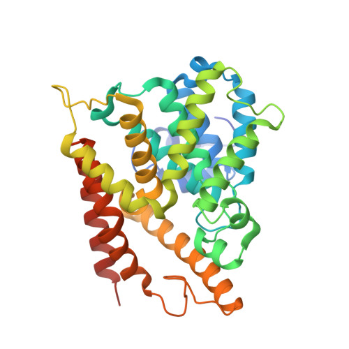
    Selected members of the large rolipram-related GEBR family of type 4 phosphodiesterase (PDE4) inhibitors have been shown to facilitate long-term potentiation and to improve memory functions without causing emetic-like behavior in rodents. Despite their micromolar-range binding affinities and their promising pharmacological and toxicological profiles, few if any structure-activity relationship studies have been performed to elucidate the molecular bases of their action. Here, we report the crystal structure of a number of GEBR library compounds in complex with the catalytic domain of PDE4D as well as their inhibitory profiles for both the long PDE4D3 isoform and the catalytic domain alone. Furthermore, we assessed the stability of the observed ligand conformations in the context of the intact enzyme using molecular dynamics simulations. The longer and more flexible ligands appear to be capable of forming contacts with the regulatory portion of the enzyme, thus possibly allowing some degree of selectivity between the different PDE4 isoforms.


  • Organizational Affiliation
    • Center for Nano Science and Technology @ PoliMi , Istituto Italiano di Tecnologia , via Giovanni Pascoli 70/3 , 20133 Milano , Italy.

Macromolecule Content 

  • Total Structure Weight: 80.42 kDa 
  • Atom Count: 5,925 
  • Modeled Residue Count: 652 
  • Deposited Residue Count: 686 
  • Unique protein chains: 1

Macromolecules

Find similar proteins by:|  3D Structure
Entity ID: 1
MoleculeChains  Sequence LengthOrganismDetailsImage
cAMP-specific 3',5'-cyclic phosphodiesterase 4D
A, B
343Homo sapiensMutation(s): 0 
Gene Names: PDE4DDPDE3
EC: 3.1.4.53
UniProt & NIH Common Fund Data Resources
Find proteins for Q08499 (Homo sapiens)
Explore Q08499 
Go to UniProtKB:  Q08499
PHAROS:  Q08499
GTEx:  ENSG00000113448 
Entity Groups
Sequence Clusters30% Identity50% Identity70% Identity90% Identity95% Identity100% Identity
UniProt GroupQ08499
Sequence Annotations
Expand
Reference Sequence

Small Molecules

Ligands 4 Unique
IDChains Name / Formula / InChI Key2D Diagram3D Interactions
CV8
(Subject of Investigation/LOI)

Query on CV8


Download:Ideal Coordinates CCD File
F [auth A],
J [auth B]
2-[(~{E})-(3-cyclopentyloxy-4-methoxy-phenyl)methylideneamino]oxy-1-[(2~{R},6~{S})-2,6-dimethylmorpholin-4-yl]ethanone
C21 H30 N2 O5
JEXHQZPMUKFOIV-RULQDBOMSA-N
GOL

Query on GOL


Download:Ideal Coordinates CCD File
G [auth A],
K [auth B]
GLYCEROL
C3 H8 O3
PEDCQBHIVMGVHV-UHFFFAOYSA-N
ZN

Query on ZN


Download:Ideal Coordinates CCD File
D [auth A],
I [auth B]
ZINC ION
Zn
PTFCDOFLOPIGGS-UHFFFAOYSA-N
MG

Query on MG


Download:Ideal Coordinates CCD File
C [auth A],
E [auth A],
H [auth B]
MAGNESIUM ION
Mg
JLVVSXFLKOJNIY-UHFFFAOYSA-N

Experimental Data & Validation

Experimental Data

  • Method: X-RAY DIFFRACTION
  • Resolution: 1.83 Å
  • R-Value Free:  0.230 (Depositor), 0.241 (DCC) 
  • R-Value Work:  0.185 (Depositor) 
  • R-Value Observed: 0.187 (Depositor) 
Space Group: P 21 21 21
Unit Cell:
Length ( Å )Angle ( ˚ )
a = 64.827α = 90
b = 98.936β = 90
c = 119.532γ = 90
Software Package:
Software NamePurpose
PHENIXrefinement
SCALAdata scaling
PHENIXphasing
XDSdata reduction

Structure Validation

View Full Validation Report


Ligand Structure Quality Assessment 


Entry History 

Deposition Data

Revision History  (Full details and data files)

  • Version 1.0: 2018-05-16
    Type: Initial release
  • Version 1.1: 2018-05-23
    Changes: Data collection, Database references
  • Version 1.2: 2024-01-17
    Changes: Data collection, Database references, Derived calculations, Refinement description