6F64 | pdb_00006f64

Crystal structure of the SYCP1 C-terminal back-to-back assembly


Experimental Data Snapshot

  • Method: X-RAY DIFFRACTION
  • Resolution: 2.49 Å
  • R-Value Free: 
    0.252 (Depositor), 0.266 (DCC) 
  • R-Value Work: 
    0.225 (Depositor), 0.244 (DCC) 
  • R-Value Observed: 
    0.227 (Depositor) 

wwPDB Validation 3D Report Full Report

Validation slider image for 6F64

This is version 1.4 of the entry. See complete history

Literature

Structural basis of meiotic chromosome synapsis through SYCP1 self-assembly.

Dunce, J.M.Dunne, O.M.Ratcliff, M.Millan, C.Madgwick, S.Uson, I.Davies, O.R.

(2018) Nat Struct Mol Biol 25: 557-569

  • DOI: https://doi.org/10.1038/s41594-018-0078-9
  • Primary Citation Related Structures: 
    6F5X, 6F62, 6F63, 6F64

  • PubMed Abstract: 

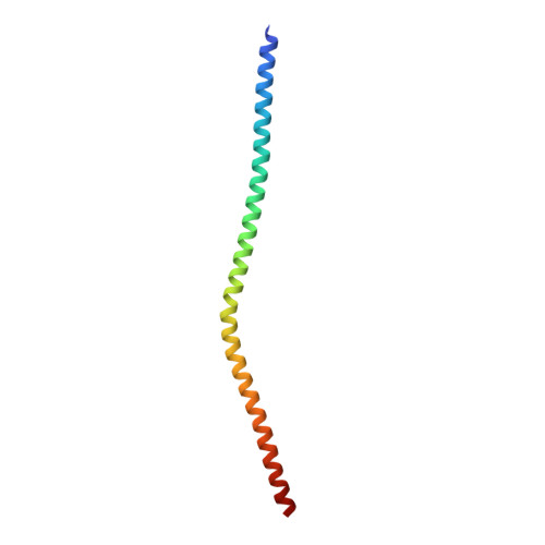
    Meiotic chromosomes adopt unique structures in which linear arrays of chromatin loops are bound together in homologous chromosome pairs by a supramolecular protein assembly, the synaptonemal complex. This three-dimensional scaffold provides the essential structural framework for genetic exchange by crossing over and subsequent homolog segregation. The core architecture of the synaptonemal complex is provided by SYCP1. Here we report the structure and self-assembly mechanism of human SYCP1 through X-ray crystallographic and biophysical studies. SYCP1 has an obligate tetrameric structure in which an N-terminal four-helical bundle bifurcates into two elongated C-terminal dimeric coiled-coils. This building block assembles into a zipper-like lattice through two self-assembly sites. N-terminal sites undergo cooperative head-to-head assembly in the midline, while C-terminal sites interact back to back on the chromosome axis. Our work reveals the underlying molecular structure of the synaptonemal complex in which SYCP1 self-assembly generates a supramolecular lattice that mediates meiotic chromosome synapsis.


  • Organizational Affiliation
    • Institute for Cell and Molecular Biosciences, Newcastle University, Newcastle upon Tyne, UK.

Macromolecule Content 

  • Total Structure Weight: 11.44 kDa 
  • Atom Count: 806 
  • Modeled Residue Count: 95 
  • Deposited Residue Count: 98 
  • Unique protein chains: 1

Macromolecules

Find similar proteins by:|  3D Structure
Entity ID: 1
MoleculeChains  Sequence LengthOrganismDetailsImage
Synaptonemal complex protein 198Homo sapiensMutation(s): 0 
Gene Names: SYCP1SCP1
UniProt & NIH Common Fund Data Resources
Find proteins for Q15431 (Homo sapiens)
Explore Q15431 
Go to UniProtKB:  Q15431
PHAROS:  Q15431
GTEx:  ENSG00000198765 
Entity Groups
Sequence Clusters30% Identity50% Identity70% Identity90% Identity95% Identity100% Identity
UniProt GroupQ15431
Sequence Annotations
Expand
Reference Sequence

Small Molecules

Ligands 1 Unique
IDChains Name / Formula / InChI Key2D Diagram3D Interactions
ACT

Query on ACT


Download:Ideal Coordinates CCD File
B [auth A]ACETATE ION
C2 H3 O2
QTBSBXVTEAMEQO-UHFFFAOYSA-M

Experimental Data & Validation

Experimental Data

  • Method: X-RAY DIFFRACTION
  • Resolution: 2.49 Å
  • R-Value Free:  0.252 (Depositor), 0.266 (DCC) 
  • R-Value Work:  0.225 (Depositor), 0.244 (DCC) 
  • R-Value Observed: 0.227 (Depositor) 
Space Group: I 41 2 2
Unit Cell:
Length ( Å )Angle ( ˚ )
a = 43.38α = 90
b = 43.38β = 90
c = 292.18γ = 90
Software Package:
Software NamePurpose
PHENIXrefinement
XDSdata reduction
XSCALEdata scaling
Arcimboldophasing

Structure Validation

View Full Validation Report


Entry History 

& Funding Information

Deposition Data


Funding OrganizationLocationGrant Number
Wellcome Trust and Royal SocietyUnited Kingdom104158/Z/14/Z

Revision History  (Full details and data files)

  • Version 1.0: 2018-06-06
    Type: Initial release
  • Version 1.1: 2018-07-04
    Changes: Data collection, Database references
  • Version 1.2: 2018-08-01
    Changes: Data collection, Database references
  • Version 1.3: 2020-04-22
    Changes: Database references
  • Version 1.4: 2024-11-13
    Changes: Data collection, Database references, Structure summary