


Nickel-Mediated Self-Assembly of an Ultrastable GB1 Variant
Bozkurt, E., Hovius, R., Browning, N.J., Perez, M.A.S., Rothlisberger, U.To be published.
Experimental Data Snapshot
Starting Model: experimental
View more details
wwPDB Validation 3D Report Full Report

Entity ID: 1 | |||||
|---|---|---|---|---|---|
| Molecule | Chains | Sequence Length | Organism | Details | Image |
| Nickel-Binding Protein | 56 | Streptococcus | Mutation(s): 0 | 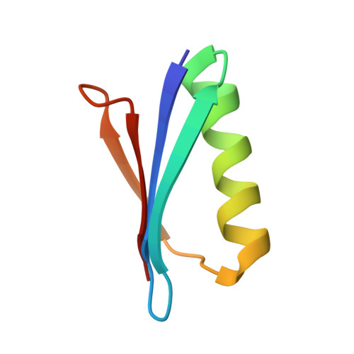 | |
| Ligands 1 Unique | |||||
|---|---|---|---|---|---|
| ID | Chains | Name / Formula / InChI Key | 2D Diagram | 3D Interactions | |
| NI (Subject of Investigation/LOI) Download:Ideal Coordinates CCD File | C [auth A] | NICKEL (II) ION Ni VEQPNABPJHWNSG-UHFFFAOYSA-N |  | ||
| Length ( Å ) | Angle ( ˚ ) |
|---|---|
| a = 26.68 | α = 90 |
| b = 59.328 | β = 90 |
| c = 75.729 | γ = 90 |
| Software Name | Purpose |
|---|---|
| PHENIX | refinement |
| ADDREF | data reduction |
| HKL-2000 | data scaling |
| PHENIX | phasing |
| Funding Organization | Location | Grant Number |
|---|---|---|
| Swiss National Science Foundation | Switzerland | 200020-165863 |


