


Crystal structure of a mutated OpuBC in complex with choline
Peherstorfer, S., Teichmann, L., Smits, S.H., Schmitt, L., Bremer, E.To be published.
Experimental Data Snapshot
Starting Model: experimental
View more details
wwPDB Validation 3D Report Full Report

Entity ID: 1 | |||||
|---|---|---|---|---|---|
| Molecule | Chains | Sequence Length | Organism | Details | Image |
| Choline-binding protein | 306 | Bacillus subtilis subsp. subtilis str. 168 | Mutation(s): 1 Gene Names: opuBC, proX, BSU33710 | 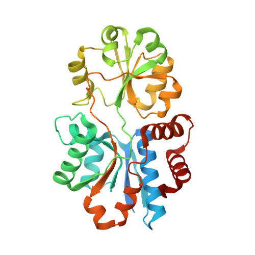 | |
UniProt | |||||
Entity Groups | |||||
| Sequence Clusters | 30% Identity50% Identity70% Identity90% Identity95% Identity100% Identity | ||||
| UniProt Group | Q45462 | ||||
Sequence AnnotationsExpand | |||||
Reference Sequence | |||||
| Ligands 1 Unique | |||||
|---|---|---|---|---|---|
| ID | Chains | Name / Formula / InChI Key | 2D Diagram | 3D Interactions | |
| CHT Download:Ideal Coordinates CCD File | C [auth A], D [auth B] | CHOLINE ION C5 H14 N O OEYIOHPDSNJKLS-UHFFFAOYSA-N |  | ||
| Length ( Å ) | Angle ( ˚ ) |
|---|---|
| a = 29.659 | α = 90 |
| b = 66.323 | β = 91.62 |
| c = 126.473 | γ = 90 |
| Software Name | Purpose |
|---|---|
| PHENIX | refinement |
| XDS | data reduction |
| XSCALE | data scaling |
| PHASER | phasing |


