Temperature-Dependent Interactions Explain Normal and Inverted Solubility in a gamma D-Crystallin Mutant.
Khan, A.R., James, S., Quinn, M.K., Altan, I., Charbonneau, P., McManus, J.J.(2019) Biophys J 117: 930-937
- PubMed: 31422822 Search on PubMedSearch on PubMed Central
- DOI: https://doi.org/10.1016/j.bpj.2019.07.019
- Primary Citation Related Structures:
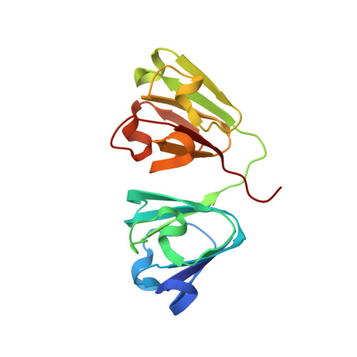
6ETA, 6ETC - PubMed Abstract:
Protein crystal production is a major bottleneck in the structural characterization of proteins. To advance beyond large-scale screening, rational strategies for protein crystallization are crucial. Understanding how chemical anisotropy (or patchiness) of the protein surface, due to the variety of amino-acid side chains in contact with solvent, contributes to protein-protein contact formation in the crystal lattice is a major obstacle to predicting and optimizing crystallization. The relative scarcity of sophisticated theoretical models that include sufficient detail to link collective behavior, captured in protein phase diagrams, and molecular-level details, determined from high-resolution structural information, is a further barrier. Here, we present two crystal structures for the P23T + R36S mutant of γD-crystallin, each with opposite solubility behavior: one melts when heated, the other when cooled. When combined with the protein phase diagram and a tailored patchy particle model, we show that a single temperature-dependent interaction is sufficient to stabilize the inverted solubility crystal. This contact, at the P23T substitution site, relates to a genetic cataract and reveals at a molecular level the origin of the lowered and retrograde solubility of the protein. Our results show that the approach employed here may present a productive strategy for the rationalization of protein crystallization.
- School of Biochemistry and Immunology, Trinity College Dublin, Dublin, Ireland.
Organizational Affiliation: