6ESZ

Crystal structure of PqsBC from Pseudomonas aeruginosa (crystal form 1)


Experimental Data Snapshot

  • Method: X-RAY DIFFRACTION
  • Resolution: 1.84 Å
  • R-Value Free: 0.179 
  • R-Value Work: 0.149 
  • R-Value Observed: 0.151 

wwPDB Validation   3D Report Full Report


This is version 1.2 of the entry. See complete history


Literature

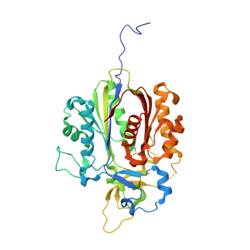
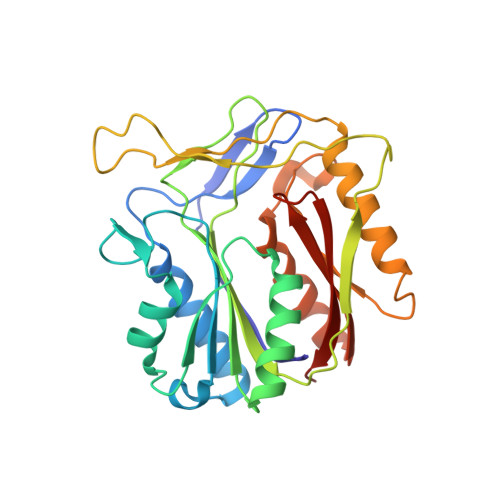
The Alkylquinolone Repertoire of Pseudomonas aeruginosa is Linked to Structural Flexibility of the FabH-like 2-Heptyl-3-hydroxy-4(1H)-quinolone (PQS) Biosynthesis Enzyme PqsBC.

Witzgall, F.Depke, T.Hoffmann, M.Empting, M.Bronstrup, M.Muller, R.Blankenfeldt, W.

(2018) Chembiochem 19: 1531-1544

  • DOI: https://doi.org/10.1002/cbic.201800153
  • Primary Citation of Related Structures:  
    6ESZ, 6ET0, 6ET1, 6ET2, 6ET3, 6ETO

  • PubMed Abstract: 

    Pseudomonas aeruginosa is a bacterial pathogen that causes life-threatening infections in immunocompromised patients. It produces a large armory of saturated and mono-unsaturated 2-alkyl-4(1H)-quinolones (AQs) and AQ N-oxides (AQNOs) that serve as signaling molecules to control the production of virulence factors and that are involved in membrane vesicle formation and iron chelation; furthermore, they also have, for example, antibiotic properties. It has been shown that the β-ketoacyl-acyl-carrier protein synthase III (FabH)-like heterodimeric enzyme PqsBC catalyzes the last step in the biosynthesis of the most abundant AQ congener, 2-heptyl-4(1H)-quinolone (HHQ), by condensing octanoyl-coenzyme A (CoA) with 2-aminobenzoylacetate (2-ABA), but the basis for the large number of other AQs/AQNOs produced by P. aeruginosa is not known. Here, we demonstrate that PqsBC uses different medium-chain acyl-CoAs to produce various saturated AQs/AQNOs and that it also biosynthesizes mono-unsaturated congeners. Further, we determined the structures of PqsBC in four different crystal forms at 1.5 to 2.7 Å resolution. Together with a previous report, the data reveal that PqsBC adopts open, intermediate, and closed conformations that alter the shape of the acyl-binding cavity and explain the promiscuity of PqsBC. The different conformations also allow us to propose a model for structural transitions that accompany the catalytic cycle of PqsBC that might have broader implications for other FabH-enzymes, for which such structural transitions have been postulated but have never been observed.


  • Organizational Affiliation

    Department Structure and Function of Proteins, Helmholtz Centre for Infection Research, Inhoffenstrasse 7, 38124, Braunschweig, Germany.


Macromolecules
Find similar proteins by:  (by identity cutoff)  |  3D Structure
Entity ID: 1
MoleculeChains Sequence LengthOrganismDetailsImage
PqsC
A, C
365Pseudomonas aeruginosa PAO1Mutation(s): 0 
Gene Names: pqsCPA0998
UniProt
Find proteins for Q9I4X1 (Pseudomonas aeruginosa (strain ATCC 15692 / DSM 22644 / CIP 104116 / JCM 14847 / LMG 12228 / 1C / PRS 101 / PAO1))
Explore Q9I4X1 
Go to UniProtKB:  Q9I4X1
Entity Groups  
Sequence Clusters30% Identity50% Identity70% Identity90% Identity95% Identity100% Identity
UniProt GroupQ9I4X1
Sequence Annotations
Expand
  • Reference Sequence
Find similar proteins by:  (by identity cutoff)  |  3D Structure
Entity ID: 2
MoleculeChains Sequence LengthOrganismDetailsImage
PqsB
B, D
283Pseudomonas aeruginosa PAO1Mutation(s): 0 
Gene Names: pqsBPA0997
UniProt
Find proteins for Q9I4X2 (Pseudomonas aeruginosa (strain ATCC 15692 / DSM 22644 / CIP 104116 / JCM 14847 / LMG 12228 / 1C / PRS 101 / PAO1))
Explore Q9I4X2 
Go to UniProtKB:  Q9I4X2
Entity Groups  
Sequence Clusters30% Identity50% Identity70% Identity90% Identity95% Identity100% Identity
UniProt GroupQ9I4X2
Sequence Annotations
Expand
  • Reference Sequence
Small Molecules
Ligands 3 Unique
IDChains Name / Formula / InChI Key2D Diagram3D Interactions
MPD
Query on MPD

Download Ideal Coordinates CCD File 
I [auth A],
J [auth A],
P [auth C],
Q [auth C],
R [auth C]
(4S)-2-METHYL-2,4-PENTANEDIOL
C6 H14 O2
SVTBMSDMJJWYQN-YFKPBYRVSA-N
PEG
Query on PEG

Download Ideal Coordinates CCD File 
K [auth A]DI(HYDROXYETHYL)ETHER
C4 H10 O3
MTHSVFCYNBDYFN-UHFFFAOYSA-N
SO4
Query on SO4

Download Ideal Coordinates CCD File 
E [auth A]
F [auth A]
G [auth A]
H [auth A]
L [auth B]
E [auth A],
F [auth A],
G [auth A],
H [auth A],
L [auth B],
M [auth B],
N [auth B],
O [auth C],
S [auth D],
T [auth D],
U [auth D]
SULFATE ION
O4 S
QAOWNCQODCNURD-UHFFFAOYSA-L
Modified Residues  1 Unique
IDChains TypeFormula2D DiagramParent
CSD
Query on CSD
A, C
L-PEPTIDE LINKINGC3 H7 N O4 SCYS
Experimental Data & Validation

Experimental Data

  • Method: X-RAY DIFFRACTION
  • Resolution: 1.84 Å
  • R-Value Free: 0.179 
  • R-Value Work: 0.149 
  • R-Value Observed: 0.151 
  • Space Group: P 21 21 21
Unit Cell:
Length ( Å )Angle ( ˚ )
a = 61.989α = 90
b = 142.384β = 90
c = 171.121γ = 90
Software Package:
Software NamePurpose
PHENIXrefinement
Aimlessdata scaling
PDB_EXTRACTdata extraction
Cootmodel building
PHASERphasing
XDSdata reduction

Structure Validation

View Full Validation Report


Entry History 

Deposition Data

Revision History  (Full details and data files)

  • Version 1.0: 2018-07-04
    Type: Initial release
  • Version 1.1: 2018-10-24
    Changes: Data collection, Database references
  • Version 1.2: 2024-01-17
    Changes: Data collection, Database references, Refinement description